 Explore
 Validate
 Learn
LearnPA1-46147
antibody from Invitrogen Antibodies
Targeting: TP53BP1
53BP1, p202, TDRD30
 Western blot Immunocytochemistry
 Immunoprecipitation Immunohistochemistry Flow cytometry Other assay
Antibody data
- Antibody Data
- Antigen structure
- References [1]
- Comments [0]
- Validations
- Western blot [4]
- Immunocytochemistry [2]
- Immunohistochemistry [1]
- Flow cytometry [3]
- Other assay [2]
Submit
Validation data
Reference
Comment
Report error
- Product number
- PA1-46147 - Provider product page

- Provider
- Invitrogen Antibodies
- Product name
- 53BP1 Polyclonal Antibody
- Antibody type
- Polyclonal
- Antigen
- Other
- Description
- Human samples have been tested in Western Blot and ICC/IF, and mouse samples have been tested in ICC/IF only. Suggested positive control: U205 cell lysates.
- Reactivity
- Human, Mouse
- Host
- Rabbit
- Isotype
- IgG
- Vial size
- 100 µL
- Concentration
- 1 mg/mL
- Storage
- -20°C
Submitted references NFATC2 Modulates Radiation Sensitivity in Dermal Fibroblasts From Patients With Severe Side Effects of Radiotherapy.
Dulong J, Kouakou C, Mesloub Y, Rorteau J, Moratille S, Chevalier FP, Vinasco-Sandoval T, Martin MT, Lamartine J
Frontiers in oncology 2020;10:589168
Frontiers in oncology 2020;10:589168
No comments: Submit comment
Supportive validation
- Submitted by
- Invitrogen Antibodies (provider)
- Main image

- Experimental details
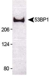
- Western blot detection of 53BP1 (220 kDa) from untreated U2OS cell lysate using Product # PA1-46147.

- Submitted by
- Invitrogen Antibodies (provider)
- Main image
- Experimental details
- Western blot analysis of 53BP1 in untreated U2OS cell lysate. Samples were incubated in 53BP1 polyclonal antibody (Product # PA1-46147). The observed molecular weight here is ~220 kDa, and the theoretical molecular weight is 214 kDa.

- Submitted by
- Invitrogen Antibodies (provider)
- Main image

- Experimental details
- Knockdown of TP53-binding protein 1 was achieved by transfecting HeLa with TP53-binding protein 1 specific siRNAs (Silencer® select Product # s14315, s14314). Western blot analysis (Fig. a) was performed using Nuclear enriched extracts from the TP53-binding protein 1 knockdown cells (lane 3), non-targeting scrambled siRNA transfected cells (lane 2) and untransfected cells (lane 1). The blot was probed with 53BP1 Polyclonal Antibody (Product # PA1-46147, 1:5000 ) and Goat anti-Rabbit IgG (H+L) Superclonal™ Recombinant Secondary Antibody, HRP (Product # A27036, 1:4000). Densitometric analysis of this western blot is shown in histogram (Fig. b). Decrease in signal upon siRNA mediated knock down confirms that antibody is specific to TP53-binding protein 1.A streak like pattern was observed in the positive cell lines as expected of 53BP1 target.

- Submitted by
- Invitrogen Antibodies (provider)
- Main image

- Experimental details
- Western blot was performed using Anti-53BP1 Polyclonal Antibody (Product # PA1-46147) and a ~270kDa band corresponding to TP53-binding protein 1 was observed across cell lines tested . Nuclear enriched extracts (30 µg lysate) of HeLa (Lane 1), A549 (Lane 2), MCF7 (Lane 3), U-2 OS (Lane 4), K-562 (Lane 5), U-87 MG (Lane 6) were electrophoresed using NuPAGE™ 3-8% Tris-Acetate Protein Gel (Product # EA0378BOX). Resolved proteins were then transferred onto a Nitrocellulose membrane (Product # LC2001) by iBlot® 2 Dry Blotting System (Product # IB21001). The blot was probed with the primary antibody (1:5000) and detected by chemiluminescence with Goat anti-Rabbit IgG (H+L) Superclonal™ Recombinant Secondary Antibody, HRP (Product # A27036, 1:4000) using the iBright FL 1000 (Product # A32752). Chemiluminescent detection was performed using Novex® ECL Chemiluminescent Substrate Reagent Kit (Product # WP20005).A streak like pattern was observed in the positive cell lines as expected of 53BP1 target.

Supportive validation
- Submitted by
- Invitrogen Antibodies (provider)
- Main image

- Experimental details
- Immunocytochemistry analysis of 53BP1 in HeLa cells fixed for 10 minutes using 10% formalin and then permeabilized for 5 minutes using 1X PBS + 0.5% Triton X-100. Samples were incubated in 53BP1 polyclonal antibody (Product # PA1-46147) using a dilution of 10 µg/mL for 1 hour at room temperature. Antibody conjugated to Alexa Fluor 488. Nuclei were counterstained with DAPI (Blue). Cells were imaged using a 40X objective.

- Submitted by
- Invitrogen Antibodies (provider)
- Main image

- Experimental details
- Immunofluorescence analysis of TP53-binding protein 1 was performed using 70% confluent log phase HeLa cells. The cells were fixed with 4% paraformaldehyde for 10 minutes, permeabilized with 0.1% Triton™ X-100 for 15 minutes, and blocked with 2% BSA for 45 minutes at room temperature. The cells were labeled with 53BP1 Polyclonal Antibody (Product # PA1-46147) at 1:500 in 0.1% BSA, incubated at 4 degree celsius overnight and then labeled with Donkey anti-Rabbit IgG (H+L) Highly Cross-Adsorbed Secondary Antibody, Alexa Fluor Plus 488 (Product # A32790), (1:2000), for 45 minutes at room temperature (Panel a: Green). Nuclei (Panel b:Blue) were stained with ProLong™ Diamond Antifade Mountant with DAPI (Product # P36962). F-actin (Panel c: Red) was stained with Rhodamine Phalloidin (Product # R415, 1:300). Panel d represents the merged image showing Nuclear localization. Panel e represents control cells with no primary antibody to assess background. The images were captured at 60X magnification.

Supportive validation
- Submitted by
- Invitrogen Antibodies (provider)
- Main image
- Experimental details
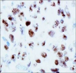
- Immunohistochemical analysis of 53BP1 in human renal cancer cells. Samples were incubated with 53BP1 polyclonal antibody (Product # PA1-46147) followed by DAB with hematoxylin counterstain.

Supportive validation
- Submitted by
- Invitrogen Antibodies (provider)
- Main image

- Experimental details
- Flow cytometry of 53BP1 in Ntera2 cells (blue) and a matched isotype control (orange). Samples were incubated in 53BP1 polyclonal antibody (Product # PA1-46147) using a dilution of 1.0 µg/mL for 30 minutes at room temperature followed by a Rabbit IgG (H+L) Cross-Adsorbed Secondary Antibody, Dylight™ 550 (Product # SA5-10033). Cells were fixed with 4% PFA and then permeabilized with 0.1% saponin.

- Submitted by
- Invitrogen Antibodies (provider)
- Main image

- Experimental details
- Flow cytometry of 53BP1 in HeLa cells. Samples were incubated in 53BP1 polyclonal antibody (Product # PA1-46147) using a dilution of 2.5 µg/mL for 30 minutes at room temperature. Antibody (blue) and a matched isotype control (orange). Cells were fixed with 4% PFA and then permeabilized with 0.1% saponin. Both antibodies were conjugated to Alexa Fluor 647.

- Submitted by
- Invitrogen Antibodies (provider)
- Main image

- Experimental details
- Flow cytometry of 53BP1 in HeLa cells. Samples were incubated in 53BP1 polyclonal antibody (Product # PA1-46147) using a dilution of 5 µg/mL for 30 minutes at room temperature. HeLa cells (blue) and a matched isotype control (orange). Cells were fixed with 4% PFA and then permeabilized with 0.1% saponin. Both antibodies were conjugated to DyLight 550.

Supportive validation
- Submitted by
- Invitrogen Antibodies (provider)
- Main image

- Experimental details
- Figure 2 Impaired DNA repair in dermal fibroblasts from overreacting patients. Immunofluorescence detection of gammaH2AX (A) and 53BP1 (B) foci, investigated 0 h, 15 min, 2, 6, and 24 h after 2 Gy irradiation in cells from patients with severe radiotherapy side-effects. The number of foci was assessed in at least 100 cells in four normal (C1, C2, C4, and C8) and four patient cell strains (P1, P6, P10, and P15). Results are mean +/- SD. The p-value was calculated by one-way ANOVA. Significant at * P < 0.05, ** P < 0.01, and **** P < 0.0001. (C) DNA damage repair ability was measured using the ExSy-SPOT chip. Fluorescence was proportional to cell ability to repair indicated DNA lesions. 8oxoG, 8-oxoGuanine; AbaS, Abasic site; CisP, Cisplatin adducts; CPD-64, Cyclobutane pyrimidine dimer - pyrimidine- (6,4)-pyrimidone photoproducts; Etheno, Etheno adducts. Results are mean +/- SD from four control fibroblast strains (C1, C2, C4, and C6) and four radiosensitive fibroblast strains (P2, P7, P8, and P10). The p-value was calculated by Student's t-test. Significant at ** P < 0.01 and **** P < 0.0001.

- Submitted by
- Invitrogen Antibodies (provider)
- Main image

- Experimental details
- Figure 6 NFATC2 downregulation leads to cellular radiosensitivity. (A) NFATC2 mRNA levels were measured by RT-qPCR in three control fibroblast strains (HNF) infected with a lentiviral vector carrying either a shRNA scramble (sh- SCR ) sequence or a shRNA targeting NFATC2 (sh- NFATC2 ). Results are mean +/- SD. The p-value was calculated using a Student's t-test. Significant at * P < 0.05. (B) Representative image of immunoblotting analysis of NFATC2 protein expression in one control cell strain (HNF A) infected with lentiviral vectors sh- SCR or sh- NFATC2 , and quantification (C) . Results are mean +/- SD from immunoblotting analysis of three different cell strains infected with lentiviral vectors sh- SCR or sh- NFATC2 . The p-value was calculated using a Student's t-test. Significant at *** P < 0.001. (D) Lower SF2 was measured by colony survival assays in cells infected with lentiviral vector sh- NFATC2 compared to cells infected with lentiviral vector sh- SCR . Results are mean +/- SD from three cell strains. The p-value was calculated using a Student's t-test. Significant at * P < 0.05. More numerous gammaH2AX (E) and 53BP1 (F) residual foci, investigated by immunofluorescence, 24 h after 2 Gy irradiation in fibroblasts infected with lentiviral vector sh- NFATC2 . Assessed in at least 100 cells in three cell strains infected with lentiviral vectors sh- SCR or sh- NFATC2 . Results are mean +/- SD. The p-value was calculated using a two-way ANOVA. Significant at **** P < 0
