Explore
 Validate
 Learn
Learn Western blot
Antibody data
- Antibody Data
- Antigen structure
- References [1]
- Comments [0]
- Validations
- Western blot [2]
- Immunocytochemistry [5]
- Immunohistochemistry [1]
Submit
Validation data
Reference
Comment
Report error
- Product number
- PA5-17578 - Provider product page

- Provider
- Invitrogen Antibodies
- Product name
- 53BP1 Polyclonal Antibody
- Antibody type
- Polyclonal
- Antigen
- Synthetic peptide
- Description
- It is not recommended to aliquot this antibody.
- Reactivity
- Human
- Host
- Rabbit
- Isotype
- IgG
- Vial size
- 100 µL
- Concentration
- 26 µg/mL
- Storage
- -20°C
Submitted references Reduced levels of methyltransferase DNMT2 sensitize human fibroblasts to oxidative stress and DNA damage that is accompanied by changes in proliferation-related miRNA expression.
Lewinska A, Adamczyk-Grochala J, Kwasniewicz E, Deregowska A, Semik E, Zabek T, Wnuk M
Redox biology 2018 Apr;14:20-34
Redox biology 2018 Apr;14:20-34
No comments: Submit comment
Supportive validation
- Submitted by
- Invitrogen Antibodies (provider)
- Main image
- Experimental details
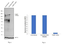
- Knockdown of TP53-binding protein 1 was achieved by transfecting HeLa with TP53-binding protein 1 specific siRNAs (Silencer® select Product # s14315, s14314). Western blot analysis (Fig. a) was performed using Nuclear enriched extracts from the TP53-binding protein 1 knockdown cells (lane 3), non-targeting scrambled siRNA transfected cells (lane 2) and untransfected cells (lane 1). The blot was probed with 53BP1 Polyclonal Antibody (Product # PA5-17578, 1:1000) and Goat anti-Rabbit IgG (H+L) Superclonal™ Recombinant Secondary Antibody, HRP (Product # A27036, 1:4000). Densitometric analysis of this western blot is shown in histogram (Fig. b). Decrease in signal upon siRNA mediated knock down confirms that antibody is specific to TP53-binding protein 1.53BP1 is reported to pick up in a streak like pattern as is seen here.

- Submitted by
- Invitrogen Antibodies (provider)
- Main image
- Experimental details
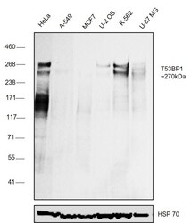
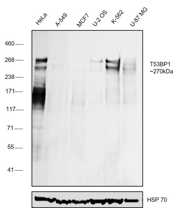
- Western blot was performed using Anti-53BP1 Polyclonal Antibody (Product # PA5-17578) and a ~270kDa band corresponding to TP53-binding protein 1 was observed across cell lines tested . Nuclear enriched extracts (30 µg lysate) of HeLa (Lane 1), A549 (Lane 2), MCF7 (Lane 3), U-2 OS (Lane 4), K-562 (Lane 5), U-87 MG (Lane 6) were electrophoresed using NuPAGE™ 3-8% Tris-Acetate Protein Gel (Product # EA0378BOX). Resolved proteins were then transferred onto a Nitrocellulose membrane (Product # LC2002) by iBlot® 2 Dry Blotting System (Product # IB21001). The blot was probed with the primary antibody (1:1000) and detected by chemiluminescence with Goat anti-Rabbit IgG (H+L) Superclonal™ Recombinant Secondary Antibody, HRP (Product # A27036, 1:4000) using the iBright FL 1000 (Product # A32752). Chemiluminescent detection was performed using Novex® ECL Chemiluminescent Substrate Reagent Kit (Product # WP20005). 53BP1 is reported to pick up as a streak like pattern which is observed in the models tested here.
Supportive validation
- Submitted by
- Invitrogen Antibodies (provider)
- Main image

- Experimental details
- Immunofluorescent analysis of 53BP1 using a polyclonal antibody (Product # PA5-17578).
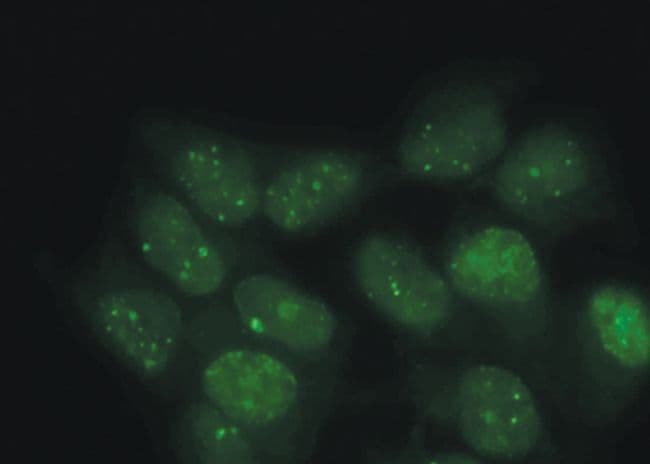
- Submitted by
- Invitrogen Antibodies (provider)
- Main image

- Experimental details
- Immunofluorescent analysis of 53BP1 using a polyclonal antibody (Product # PA5-17578).

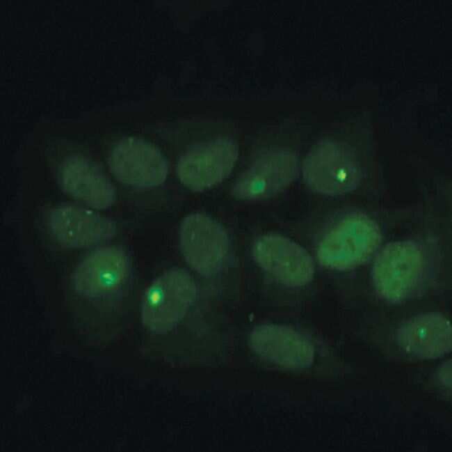
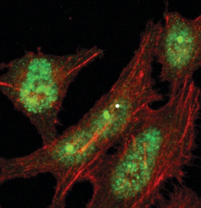
- Submitted by
- Invitrogen Antibodies (provider)
- Main image

- Experimental details
- Immunofluorescent analysis of 53BP1 in HeLa cells using a 53BP1 polyclonal antibody (Product # PA5-17578) (green). Actin filaments are labeled with a fluorescent red phalloidin.
- Submitted by
- Invitrogen Antibodies (provider)
- Main image

- Experimental details
- Immunofluorescent analysis of 53BP1 using a polyclonal antibody (Product # PA5-17578).
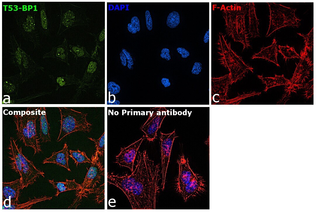
- Submitted by
- Invitrogen Antibodies (provider)
- Main image

- Experimental details
- Immunofluorescence analysis of TP53-binding protein 1 was performed using 70% confluent log phase HeLa cells. The cells were fixed with 4% paraformaldehyde for 10 minutes, permeabilized with 0.1% Triton™ X-100 for 15 minutes, and blocked with 2% BSA for 45 minutes at room temperature. The cells were labeled with 53BP1 Polyclonal Antibody (Product # PA5-17578) at 1:100 in 0.1% BSA, incubated at 4 degree celsius overnight and then labeled with Donkey anti-Rabbit IgG (H+L) Highly Cross-Adsorbed Secondary Antibody, Alexa Fluor Plus 488 (Product # A32790), (1:2000), for 45 minutes at room temperature (Panel a: Blue). Nuclei (Panel b:Green) were stained with ProLong™ Diamond Antifade Mountant with DAPI (Product # P36962). F-actin (Panel c: Red) was stained with Rhodamine Phalloidin (Product # R415, 1:300). Panel d represents the merged image showing Nuclear localization. Panel e represents control cells with no primary antibody to assess background. The images were captured at 60X magnification.
Supportive validation
- Submitted by
- Invitrogen Antibodies (provider)
- Main image
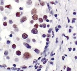
- Experimental details
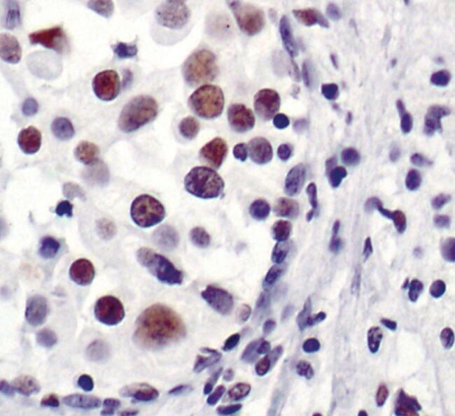
- Immunohistochemical analysis of 53BP1 in paraffin-embedded human breast carcinoma using a 53BP1 polyclonal antibody (Product # PA5-17578) showing nuclear localization.