Explore
 Validate
 Learn
Learn Western blot
 Immunocytochemistry
Antibody data
- Antibody Data
- Antigen structure
- References [1]
- Comments [0]
- Validations
- Immunocytochemistry [4]
- Immunohistochemistry [3]
- Flow cytometry [3]
- Other assay [1]
Submit
Validation data
Reference
Comment
Report error
- Product number
- MA5-32687 - Provider product page

- Provider
- Invitrogen Antibodies
- Product name
- MRP2 Recombinant Rabbit Monoclonal Antibody (JA32-01)
- Antibody type
- Monoclonal
- Antigen
- Synthetic peptide
- Description
- Recombinant rabbit monoclonal antibodies are produced using in vitro expression systems. The expression systems are developed by cloning in the specific antibody DNA sequences from immunoreactive rabbits. Then, individual clones are screened to select the best candidates for production. The advantages of using recombinant rabbit monoclonal antibodies include: better specificity and sensitivity, lot-to-lot consistency, animal origin-free formulations, and broader immunoreactivity to diverse targets due to larger rabbit immune repertoire.
- Reactivity
- Human
- Host
- Rabbit
- Isotype
- IgG
- Antibody clone number
- JA32-01
- Vial size
- 100 μL
- Concentration
- 1 mg/mL
- Storage
- Store at 4°C short term. For long term storage, store at -20°C, avoiding freeze/thaw cycles.
Submitted references Generation of Human Pluripotent Stem Cell-Derived Polarized Hepatocytes.
Bushweller L, Zhao Y, Zhang F, Wu X
Current protocols 2022 Jan;2(1):e345
Current protocols 2022 Jan;2(1):e345
No comments: Submit comment
Supportive validation
- Submitted by
- Invitrogen Antibodies (provider)
- Main image

- Experimental details
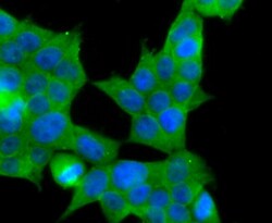
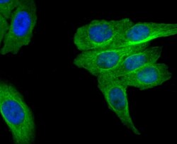
- Immunocytochemical analysis of MRP2 in HepG2 cells using a MRP2 Monoclonal antibody (Product # MA5-32687) as seen in green. The nuclear counter stain is DAPI (blue). Cells were fixed in paraformaldehyde, permeabilised with 0.25% Triton X100/PBS.

- Submitted by
- Invitrogen Antibodies (provider)
- Main image
- Experimental details
- Immunocytochemical analysis of MRP2 in 293T cells using a MRP2 Monoclonal antibody (Product # MA5-32687) as seen in green. The nuclear counter stain is DAPI (blue). Cells were fixed in paraformaldehyde, permeabilised with 0.25% Triton X100/PBS.

- Submitted by
- Invitrogen Antibodies (provider)
- Main image
- Experimental details
- Immunocytochemical analysis of MRP2 in HepG2 cells using a MRP2 Monoclonal antibody (Product # MA5-32687) as seen in green. The nuclear counter stain is DAPI (blue). Cells were fixed in paraformaldehyde, permeabilised with 0.25% Triton X100/PBS.

- Submitted by
- Invitrogen Antibodies (provider)
- Main image

- Experimental details
- Immunocytochemical analysis of MRP2 in 293T cells using a MRP2 Monoclonal antibody (Product # MA5-32687) as seen in green. The nuclear counter stain is DAPI (blue). Cells were fixed in paraformaldehyde, permeabilised with 0.25% Triton X100/PBS.

Supportive validation
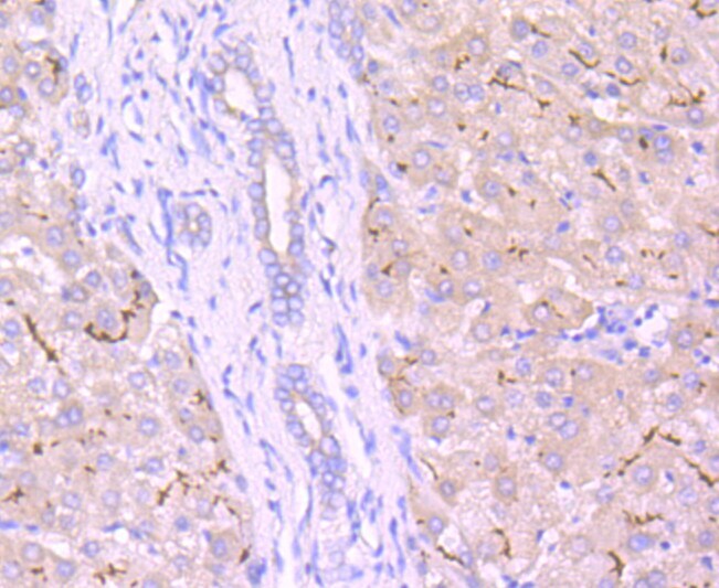
- Submitted by
- Invitrogen Antibodies (provider)
- Main image
- Experimental details
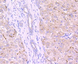
- Immunohistochemical analysis of MRP2 of paraffin-embedded Human liver tissue using a MRP2 Monoclonal antibody (Product #MA5-32687). Counter stained with hematoxylin.
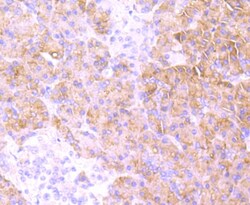
- Submitted by
- Invitrogen Antibodies (provider)
- Main image
- Experimental details
- Immunohistochemical analysis of MRP2 of paraffin-embedded Human kidney tissue using a MRP2 Monoclonal antibody (Product #MA5-32687). Counter stained with hematoxylin.

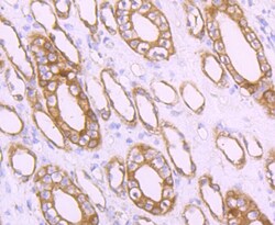
- Submitted by
- Invitrogen Antibodies (provider)
- Main image
- Experimental details
- Immunohistochemical analysis of MRP2 of paraffin-embedded Human pancreas tissue using a MRP2 Monoclonal antibody (Product #MA5-32687). Counter stained with hematoxylin.

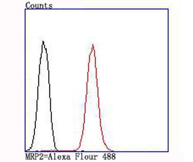
Supportive validation
- Submitted by
- Invitrogen Antibodies (provider)
- Main image
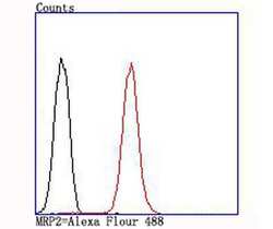
- Experimental details
- Flow Cytometric analysis of MRP2 in A549 cells using a MRP2 Monoclonal Antibody (Product # MA5-32687) at a dilution of 1:100, as seen in red compared with an unlabelled control (cells without incubation with primary antibody; black).
- Submitted by
- Invitrogen Antibodies (provider)
- Main image
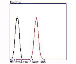
- Experimental details
- Flow Cytometric analysis of MRP2 in A549 cells using a MRP2 Monoclonal Antibody (Product # MA5-32687) at a dilution of 1:100, as seen in red compared with an unlabelled control (cells without incubation with primary antibody; black).
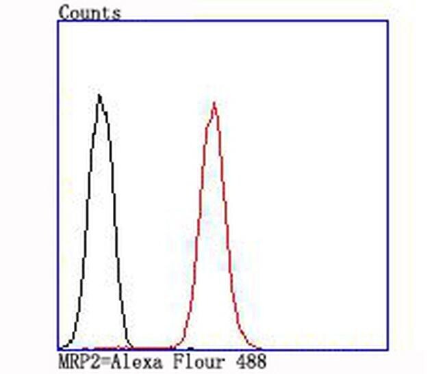
Supportive validation
- Submitted by
- Invitrogen Antibodies (provider)
- Main image
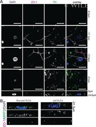
- Experimental details
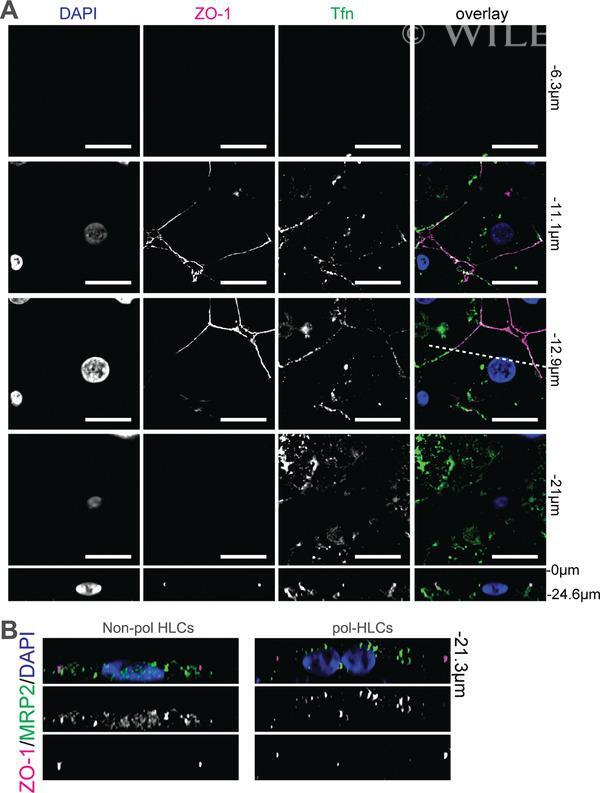
- 4 Figure Distribution of membrane proteins TFRC and MRP2 on pol-HLCs. ( A ) pol-HLCs were incubated with 25 ug/ml of transferrin-488 (green) for 10 min at 37degC prior to washing and staining with anti-ZO-1 (red). Scale bars = 15 mum. Bottom panels: xz images from cross sections indicated by the dashed line in the corresponding xy images above. ( B ) Cross-sectional views ( xz ) of nonpolarized HLCs (non-pol HLCs) and pol-HLCs stained for MRP2 and ZO-1. This figure is adapted from Dao Thi et al. ( 2020).