 Explore
 Validate
 Learn
Learn Western blot
 Immunocytochemistry
Antibody data
- Antibody Data
- Antigen structure
- References [1]
- Comments [0]
- Validations
- Western blot [1]
Submit
Validation data
Reference
Comment
Report error
- Product number
- GTX25246 - Provider product page

- Provider
- GeneTex
- Proper citation
- GeneTex Cat#GTX25246, RRID:AB_380487
- Product name
- NFAT1 (phospho Ser54) antibody
- Antibody type
- Polyclonal
- Reactivity
- Human, Mouse
- Host
- Rabbit
Submitted references 6-Methoxyflavone inhibits NFAT translocation into the nucleus and suppresses T cell activation.
So JS, Kim GC, Song M, Lee CG, Park E, Kim HJ, Kim YS, Jun CD, Im SH
Journal of immunology (Baltimore, Md. : 1950) 2014 Sep 15;193(6):2772-83
Journal of immunology (Baltimore, Md. : 1950) 2014 Sep 15;193(6):2772-83
No comments: Submit comment
Supportive validation
- Submitted by
- GeneTex (provider)
- Main image
- Experimental details
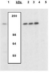
- Extracts of Murine T cells were left untreated (1) or treated with PMA and Ca2+ ionophore ionomycin (2-5), were resolved by SDS-PAGE on a 10% Tris-glycine gel and transferred to PVDF. The membrane was blocked with a 5% BSA-TBST buffer overnight at 4 degree C, then incubated with the NFAT1 [pS54] (mouse) antibody in a 3% BSA-TBST buffer for two hours at room temperature, following prior incubation with: no peptide (1,2), the non-phosphopeptide corresponding to the phosphopeptide immunogen (3), a generic phosphoserine-containing peptide (4), or the phosphopeptide immunogen (5). After washing, the membrane was incubated with goat anti rabbit IgG alkaline phosphatase. The data show only the phopshopeptide corresponding to NFAT1 [pS54] (mouse) completely blocks the antibody signal, demonstrating specificity of the antibody. The data also show the upregulation of NFAT1 upon PMA Ca2+ ionophore ionomycin treatment in this cell system.
