Explore
 Validate
 Learn
LearnGTX22722
antibody from GeneTex
Targeting: NFATC2
NF-ATP, NFAT1, NFATp
 Western blot
 Immunocytochemistry Immunoprecipitation Immunohistochemistry Flow cytometry Gel shift Chromatin Immunoprecipitation
Antibody data
- Antibody Data
- Antigen structure
- References [1]
- Comments [0]
- Validations
- Immunocytochemistry [4]
- Immunohistochemistry [3]
Submit
Validation data
Reference
Comment
Report error
- Product number
- GTX22722 - Provider product page

- Provider
- GeneTex
- Proper citation
- GeneTex Cat#GTX22722, RRID:AB_372490
- Product name
- NFAT1 antibody [25A10.D6.D2]
- Antibody type
- Monoclonal
- Reactivity
- Human, Mouse, Rat
- Host
- Mouse
Submitted references CD5 promotes IL-10 production in chronic lymphocytic leukemia B cells through STAT3 and NFAT2 activation.
Garaud S, Morva A, Lemoine S, Hillion S, Bordron A, Pers JO, Berthou C, Mageed RA, Renaudineau Y, Youinou P
Journal of immunology (Baltimore, Md. : 1950) 2011 Apr 15;186(8):4835-44
Journal of immunology (Baltimore, Md. : 1950) 2011 Apr 15;186(8):4835-44
No comments: Submit comment
Supportive validation
- Submitted by
- GeneTex (provider)
- Main image
- Experimental details
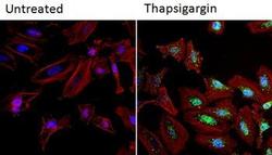
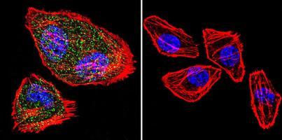
- Immunofluorescent analysis of NFATc2 (green) in HeLa cells. Formalin fixed cells were permeabilized with 0.1% Triton X-100 in TBS for 10 minutes at room temperature and blocked with 1% Blocker BSA for 15 minutes at room temperature. Cells were left untreated (left panel) or treated with 1uM staurosporine (right panel) for 3 hours and probed with a NFATc2 monoclonal antibody (GTX22722), at a dilution of 1:100 for at least 1 hour at room temperature, washed with PBS, and incubated with DyLight 488 goat anti-mouse IgG secondary antibody at a dilution of 1:400 for 30 minutes at room temperature. F-Actin (red) was stained with DyLight 554 Phalloidin and nuclei (blue) were stained with Hoechst 33342 dye.

- Submitted by
- GeneTex (provider)
- Main image

- Experimental details
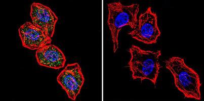
- Immunofluorescent analysis of NFATc2 using NFATc2 Monoclonal Antibody (GTX22722) shows staining in Hela Cells. NFATc2 (green), F-Actin staining with Phalloidin (red) and nuclei with DAPI (blue) is shown. Cells were grown on chamber slides and fixed with formaldehyde prior to staining. Cells were probed without (control) or with an antibody recognizing NFATc2 (GTX22722) at a dilution of 1:20 overnight at 4 degree C, washed with PBS and incubated with a DyLight-488 conjugated secondary antibody.
- Submitted by
- GeneTex (provider)
- Main image

- Experimental details
- Immunofluorescent analysis of NFATc2 using NFATc2 monoclonal antibody (GTX22722) shows staining in MCF-7 Cells. NFATc2 (green), F-Actin staining with Phalloidin (red) and nuclei with DAPI (blue) is shown. Cells were grown on chamber slides and fixed with formaldehyde prior to staining. Cells were probed without (control) or with an antibody recognizing NFATc2 (GTX22722) at a dilution of 1:20 over night at 4 C, washed with PBS and incubated with a DyLight-488 conjugated secondary antibody.
- Submitted by
- GeneTex (provider)
- Main image

- Experimental details
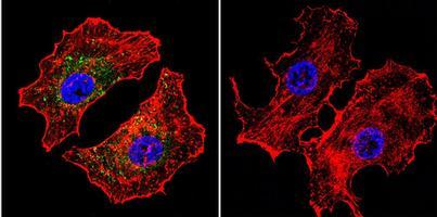
- Immunofluorescent analysis of NFATc2 using NFATc2 monoclonal antibody (GTX22722) shows staining in U251 Cells. NFATc2 (green), F-Actin staining with Phalloidin (red) and nuclei with DAPI (blue) is shown. Cells were grown on chamber slides and fixed with formaldehyde prior to staining. Cells were probed without (control) or with an antibody recognizing NFATc2 (GTX22722) at a dilution of 1:20 over night at 4 degree C, washed with PBS and incubated with a DyLight-488 conjugated secondary antibody.
Supportive validation
- Submitted by
- GeneTex (provider)
- Main image
- Experimental details
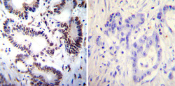
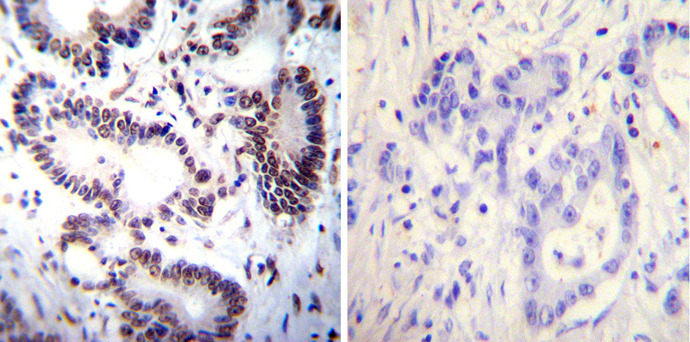
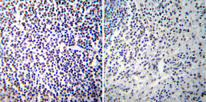
- Immunohistochemistry was performed on cancer biopsies of deparaffinized Human colon carcinoma tissues. To expose target proteins, heat induced antigen retrieval was performed using 10mM sodium citrate (pH6.0) buffer, microwaved for 8-15 minutes. Following antigen retrieval tissues were blocked in 3% BSA-PBS for 30 minutes at room temperature. Tissues were then probed at a dilution of 1:100 with a mouse monoclonal antibody recognizing NFATc2 (MA1-025) or without primary antibody (negative control) overnight at 4¢XC in a humidified chamber. Tissues were washed extensively with PBST and endogenous peroxidase activity was quenched with a peroxidase suppressor. Detection was performed using a biotin-conjugated secondary antibody and SA-HRP, followed by colorimetric detection using DAB. Tissues were counterstained with hematoxylin and prepped for mounting.
- Submitted by
- GeneTex (provider)
- Main image
- Experimental details
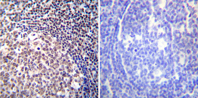
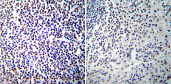
- Immunohistochemistry was performed on normal de-paraffinized human spleen tissue tissues. To expose target proteins, heat induced antigen retrieval was performed using 10mM sodium citrate (pH6.0) buffer, microwaved for 8-15 minutes. Following antigen retrieval tissues were blocked in 3% BSA-PBS for 30 minutes at room temperature. Tissues were then probed at a dilution of 1:100 with a mouse monoclonal antibody recognizing NFATc2 (GTX22722) or without primary antibody (negative control) overnight at 4¢XC in a humidified chamber. Tissues were washed extensively with PBST and endogenous peroxidase activity was quenched with a peroxidase suppressor. Detection was performed using a biotin-conjugated secondary antibody and SA-HRP, followed by colorimetric detection using DAB. Tissues were counterstained with hematoxylin and prepped for mounting.
- Submitted by
- GeneTex (provider)
- Main image
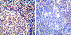
- Experimental details
- Immunohistochemistry was performed on normal de-paraffinized human tonsil tissue tissues. To expose target proteins, heat induced antigen retrieval was performed using 10mM sodium citrate (pH6.0) buffer, microwaved for 8-15 minutes. Following antigen retrieval tissues were blocked in 3% BSA-PBS for 30 minutes at room temperature. Tissues were then probed at a dilution of 1:100 with a mouse monoclonal antibody recognizing NFATc2 (GTX22722) or without primary antibody (negative control) overnight at 4¢XC in a humidified chamber. Tissues were washed extensively with PBST and endogenous peroxidase activity was quenched with a peroxidase suppressor. Detection was performed using a biotin-conjugated secondary antibody and SA-HRP, followed by colorimetric detection using DAB. Tissues were counterstained with hematoxylin and prepped for mounting.