Explore
 Validate
 Learn
Learn Western blot
 Immunocytochemistry
Antibody data
- Antibody Data
- Antigen structure
- References [5]
- Comments [0]
- Validations
- Immunocytochemistry [1]
- Immunohistochemistry [1]
Submit
Validation data
Reference
Comment
Report error
- Product number
- HPA008789 - Provider product page

- Provider
- Atlas Antibodies
- Proper citation
- Atlas Antibodies Cat#HPA008789, RRID:AB_1079474
- Product name
- Anti-NFATC2
- Antibody type
- Polyclonal
- Description
- Polyclonal Antibody against Human NFATC2, Gene description: nuclear factor of activated T-cells, cytoplasmic, calcineurin-dependent 2, Alternative Gene Names: NF-ATP, NFAT1, NFATp, Validated applications: WB, IHC, ICC, Uniprot ID: Q13469, Storage: Store at +4°C for short term storage. Long time storage is recommended at -20°C.
- Reactivity
- Human
- Host
- Rabbit
- Conjugate
- Unconjugated
- Isotype
- IgG
- Vial size
- 100 µl
- Concentration
- 0.1 mg/ml
- Storage
- Store at +4°C for short term storage. Long time storage is recommended at -20°C.
- Handling
- The antibody solution should be gently mixed before use.
Submitted references Myeloid antigen-presenting cell niches sustain antitumor T cells and license PD-1 blockade via CD28 costimulation
TcellSubC: An Atlas of the Subcellular Proteome of Human T Cells.
Nfat/calcineurin signaling promotes oligodendrocyte differentiation and myelination by transcription factor network tuning
The calcineurin/NFAT pathway is activated in diagnostic breast cancer cases and is essential to survival and metastasis of mammary cancer cells
Galanin modulates the neural niche to favour perineural invasion in head and neck cancer
Duraiswamy J, Turrini R, Minasyan A, Barras D, Crespo I, Grimm A, Casado J, Genolet R, Benedetti F, Wicky A, Ioannidou K, Castro W, Neal C, Moriot A, Renaud-Tissot S, Anstett V, Fahr N, Tanyi J, Eiva M, Jacobson C, Montone K, Westergaard M, Svane I, Kandalaft L, Delorenzi M, Sorger P, Färkkilä A, Michielin O, Zoete V, Carmona S, Foukas P, Powell D, Rusakiewicz S, Doucey M, Dangaj Laniti D, Coukos G
Cancer Cell 2021;39(12):1623-1642.e20
Cancer Cell 2021;39(12):1623-1642.e20
TcellSubC: An Atlas of the Subcellular Proteome of Human T Cells.
Joshi RN, Stadler C, Lehmann R, Lehtiö J, Tegnér J, Schmidt A, Vesterlund M
Frontiers in immunology 2019;10:2708
Frontiers in immunology 2019;10:2708
Nfat/calcineurin signaling promotes oligodendrocyte differentiation and myelination by transcription factor network tuning
Weider M, Starost L, Groll K, Küspert M, Sock E, Wedel M, Fröb F, Schmitt C, Baroti T, Hartwig A, Hillgärtner S, Piefke S, Fadler T, Ehrlich M, Ehlert C, Stehling M, Albrecht S, Jabali A, Schöler H, Winkler J, Kuhlmann T, Wegner M
Nature Communications 2018;9(1)
Nature Communications 2018;9(1)
The calcineurin/NFAT pathway is activated in diagnostic breast cancer cases and is essential to survival and metastasis of mammary cancer cells
Tran Quang C, Leboucher S, Passaro D, Fuhrmann L, Nourieh M, Vincent-Salomon A, Ghysdael J
Cell Death & Disease 2015;6(2):e1658-e1658
Cell Death & Disease 2015;6(2):e1658-e1658
Galanin modulates the neural niche to favour perineural invasion in head and neck cancer
Scanlon C, Banerjee R, Inglehart R, Liu M, Russo N, Hariharan A, van Tubergen E, Corson S, Asangani I, Mistretta C, Chinnaiyan A, D’Silva N
Nature Communications 2015;6(1)
Nature Communications 2015;6(1)
No comments: Submit comment
Supportive validation
- Submitted by
- Atlas Antibodies (provider)
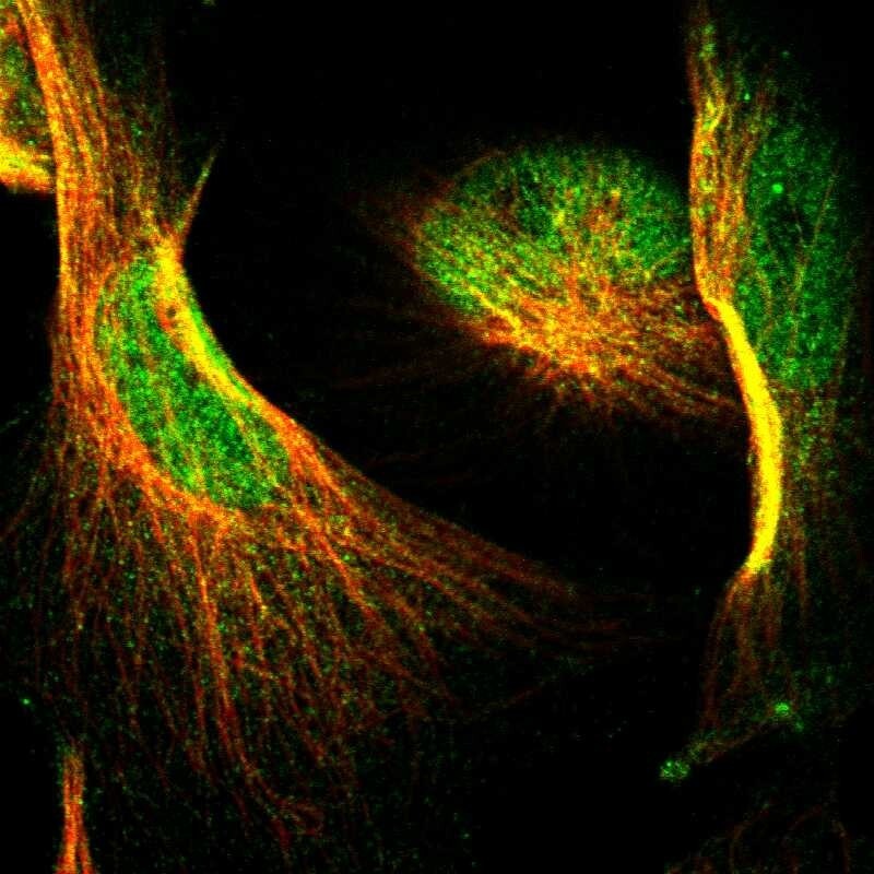
- Main image

- Experimental details
- Immunofluorescent staining of human cell line U-251 MG shows localization to nucleoplasm & cytosol.
- Sample type
- Human
Supportive validation
- Submitted by
- Atlas Antibodies (provider)
- Enhanced method
- Orthogonal validation
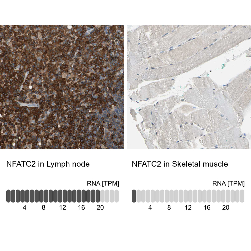
- Main image
- Experimental details
- Immunohistochemistry analysis in human lymph node and skeletal muscle tissues using HPA008789 antibody. Corresponding NFATC2 RNA-seq data are presented for the same tissues.
- Sample type
- Human
- Protocol
- Protocol