Explore
 Validate
 Learn
Learn Western blot
 Immunocytochemistry
Antibody data
- Antibody Data
- Antigen structure
- References [0]
- Comments [0]
- Validations
- Immunocytochemistry [2]
- Other assay [2]
Submit
Validation data
Reference
Comment
Report error
- Product number
- PA5-47359 - Provider product page

- Provider
- Invitrogen Antibodies
- Product name
- IL1R2 Polyclonal Antibody
- Antibody type
- Polyclonal
- Antigen
- Recombinant full-length protein
- Description
- In direct ELISAs, less than 1% cross-reactivity with recombinant human (rh) IL-1 alpha, rhIL-1 RI, recombinant mouse (rm) IL-1 alpha, rhIL-1 beta, rmIL-1 beta, recombinant rat IL-1 beta, rhIL-1ra, rmIL-1ra, and recombinant rhesus monkey IL-1ra is observed. Reconstitute at 0.2 mg/mL in sterile PBS. Endoxin level is
- Reactivity
- Human
- Host
- Goat
- Isotype
- IgG
- Vial size
- 100 μg
- Concentration
- 0.2 mg/mL
- Storage
- -20°C, Avoid Freeze/Thaw Cycles
No comments: Submit comment
Supportive validation
- Submitted by
- Invitrogen Antibodies (provider)
- Main image

- Experimental details
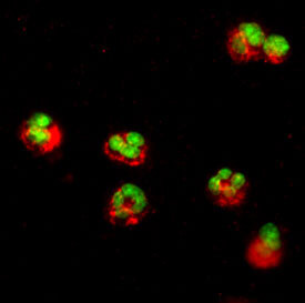
- Immunocytochemistry analysis of IL1R2 in immersion fixed human peripheral blood mononuclear cells (PBMCs). Samples were incubated in IL1R2 polyclonal antibody (Product # PA5-47359) using a dilution of 15 µg/mL for 3 hours at room temperature. Cells were stained (red) and counterstained (green).

- Submitted by
- Invitrogen Antibodies (provider)
- Main image

- Experimental details
- Immunocytochemistry analysis of IL1R2 in immersion fixed human peripheral blood mononuclear cells (PBMCs). Samples were incubated in IL1R2 polyclonal antibody (Product # PA5-47359) using a dilution of 15 µg/mL for 3 hours at room temperature. Cells were stained (red) and counterstained (green).
Supportive validation
- Submitted by
- Invitrogen Antibodies (provider)
- Main image

- Experimental details
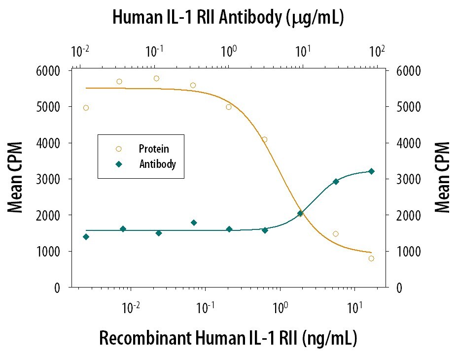
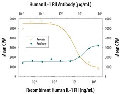
- Neutralization of IL1R2 in D10.G4.1 mouse helper T cell line. Samples were incubated in IL1R2 polyclonal antibody (Product # PA5-47359). Recombinant Human IL‚1 RII inhibits Recombinant Human IL‚1β/IL‚1F2 induced proliferation in the D10.G4.1 mouse helper T cell line in a dose-dependent manner (orange line). Inhibition of Recombinant Human IL‚1β/IL‚1F2 (50 pg/mL) activity elicited by Recombinant Human IL‚1 RII (2 µg/mL) is neutralized (green line) by increasing concentrations of Goat Anti-Human IL‚1 RII Antigen Affinity-purified Polyclonal Antibody. The ND50 is typically 15‚30 µg/mL.

- Submitted by
- Invitrogen Antibodies (provider)
- Main image
- Experimental details
- Neutralization of IL1R2 in D10.G4.1 mouse helper T cell line. Samples were incubated in IL1R2 polyclonal antibody (Product # PA5-47359). Recombinant Human IL‚1 RII inhibits Recombinant Human IL‚1β/IL‚1F2 induced proliferation in the D10.G4.1 mouse helper T cell line in a dose-dependent manner (orange line). Inhibition of Recombinant Human IL‚1β/IL‚1F2 (50 pg/mL) activity elicited by Recombinant Human IL‚1 RII (2 µg/mL) is neutralized (green line) by increasing concentrations of Goat Anti-Human IL‚1 RII Antigen Affinity-purified Polyclonal Antibody. The ND50 is typically 15‚30 µg/mL.