 Explore
 Validate
 Learn
Learn Western blot
 Immunocytochemistry
Antibody data
- Antibody Data
- Antigen structure
- References [2]
- Comments [0]
- Validations
- Western blot [2]
- Immunohistochemistry [3]
- Chromatin Immunoprecipitation [1]
Submit
Validation data
Reference
Comment
Report error
- Product number
- NBP1-32681 - Provider product page

- Provider
- Novus Biologicals
- Proper citation
- Novus Cat#NBP1-32681, RRID:AB_2125179
- Product name
- Rabbit Polyclonal IL-1 RII Antibody
- Antibody type
- Polyclonal
- Description
- Immunogen affinity purified.
- Reactivity
- Human, Mouse, Rat
- Host
- Rabbit
- Isotype
- IgG
- Vial size
- 100 ul
- Storage
- Aliquot and store at -20C or -80C. Avoid freeze-thaw cycles.
Submitted references TNF-α antagonist attenuates systemic lipopolysaccharide-induced brain white matter injury in neonatal rats.
Characteristic immune, apoptosis and inflammatory gene profiles associated with intestinal acute cellular rejection in formalin-fixed paraffin-embedded mucosal biopsies.
Shin SH, Kim EK, Lee KY, Kim HS
BMC neuroscience 2019 Aug 30;20(1):45
BMC neuroscience 2019 Aug 30;20(1):45
Characteristic immune, apoptosis and inflammatory gene profiles associated with intestinal acute cellular rejection in formalin-fixed paraffin-embedded mucosal biopsies.
Asaoka T, Island ER, Tryphonopoulos P, Selvaggi G, Moon J, Tekin A, Amador A, Levi DM, Garcia J, Smith L, Nishida S, Weppler D, Tzakis AG, Ruiz P
Transplant international : official journal of the European Society for Organ Transplantation 2011 Jul;24(7):697-707
Transplant international : official journal of the European Society for Organ Transplantation 2011 Jul;24(7):697-707
No comments: Submit comment
Supportive validation
- Submitted by
- Novus Biologicals (provider)
- Main image

- Experimental details
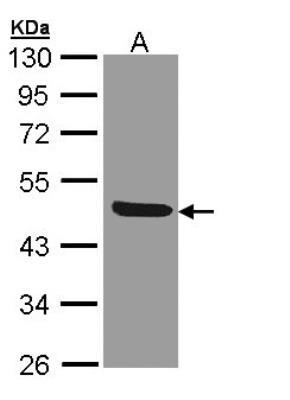
- Western Blot: IL-1 RII Antibody [NBP1-32681] - Sample (30 ug of whole cell lysate)A: 293T 10% SDS PAGE, antibody diluted at 1:1000.
- Submitted by
- Novus Biologicals (provider)
- Main image
- Experimental details
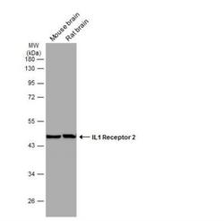
- Western Blot: IL-1 RII Antibody [NBP1-32681] - Various tissue extracts (50 ug) were separated by 10% SDS-PAGE, and the membrane was blotted with IL1 Receptor 2 antibody [N3C3] diluted at 1:1000. The HRP-conjugated anti-rabbit IgG antibody (NBP2-19301) was used to detect the primary antibody.
Supportive validation
- Submitted by
- Novus Biologicals (provider)
- Main image
- Experimental details
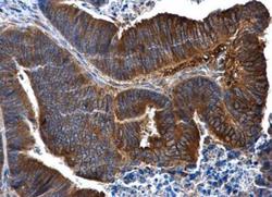
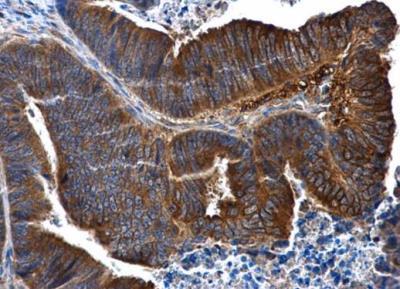
- Immunohistochemistry-Paraffin: IL-1 RII Antibody [NBP1-32681] - Paraffin-embedded human colon cancer. IL1 Receptor 2 antibody [N3C3] diluted at 1:500.
- Submitted by
- Novus Biologicals (provider)
- Main image
- Experimental details
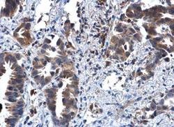
- Immunohistochemistry-Paraffin: IL-1 RII Antibody [NBP1-32681] - Paraffin-embedded human lung cancer. IL1 Receptor 2 antibody [N3C3] diluted at 1:500.

- Submitted by
- Novus Biologicals (provider)
- Main image
- Experimental details
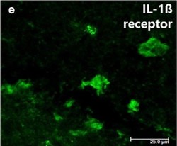
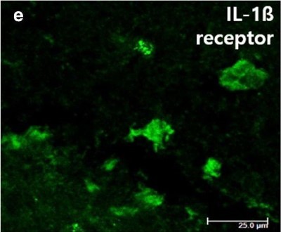
- Immunohistochemistry: IL-1 RII Antibody [NBP1-32681] - Representative photomicrographs of IL-1beta receptor immunofluorescence staining of the cingulum (Bregma - 1.0) on P4. Image collected and cropped by CiteAb from the following publication (https://bmcneurosci.biomedcentral.com/articles/10.1186/s12868-019-0529-1), licensed under a CC-BY licence.
Supportive validation
- Submitted by
- Novus Biologicals (provider)
- Main image
- Experimental details
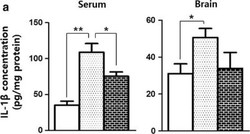
- Chromatin Immunoprecipitation: IL-1 RII Antibody [NBP1-32681] - The LPS-induced changes in the levels of inflammatory cytokines in circulation and the brain tissue of rats on P4 (IL-1beta ). ** and *represent significant differences between groups (P < 0.01 and P < 0.05, respectively). ANOVA with a Bonferroni test was conducted. There were six rats per group Image collected and cropped by CiteAb from the following publication (https://bmcneurosci.biomedcentral.com/articles/10.1186/s12868-019-0529-1), licensed under a CC-BY licence.
