Explore
 Validate
 Learn
Learn Western blot
 Immunocytochemistry
Antibody data
- Antibody Data
- Antigen structure
- References [1]
- Comments [0]
- Validations
- Immunocytochemistry [3]
- Immunohistochemistry [1]
- Other assay [3]
Submit
Validation data
Reference
Comment
Report error
- Product number
- PA5-106093 - Provider product page

- Provider
- Invitrogen Antibodies
- Product name
- Phospho-IRF5 (Ser437) Polyclonal Antibody
- Antibody type
- Polyclonal
- Antigen
- Synthetic peptide
- Description
- Antibody detects endogenous levels of IRF5 only when phosphorylated at Ser437.
- Reactivity
- Human, Mouse, Rat
- Host
- Rabbit
- Isotype
- IgG
- Vial size
- 100 μL
- Concentration
- 1 mg/mL
- Storage
- -20°C
Submitted references Phosphorylation of Microglial IRF5 and IRF4 by IRAK4 Regulates Inflammatory Responses to Ischemia.
Ngwa C, Mamun AA, Xu Y, Sharmeen R, Liu F
Cells 2021 Jan 30;10(2)
Cells 2021 Jan 30;10(2)
No comments: Submit comment
Supportive validation
- Submitted by
- Invitrogen Antibodies (provider)
- Main image
- Experimental details
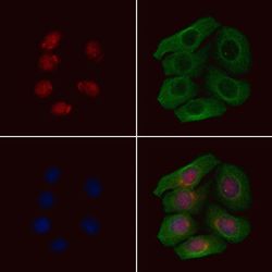
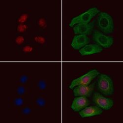
- Immunofluorescent analysis of Phospho-IRF5 (Ser437) in HeLa cells(4h of LPS treatment). Samples were fixed with paraformaldehyde, permeabilized with 0.1% Triton X-100, blocked with 10% serum (45 min at 25°C), incubated with mouse anti-beta tubulin and Phospho-IRF5 (Ser437) polyclonal antibody (Product # PA5-106093) using a dilution of 1:200 (1 hr, 37°C), and followed by goat anti-rabbit IgG Alexa Fluor 594 (red) and goat anti-mouse IgG Alexa Fluor 488 (green).

- Submitted by
- Invitrogen Antibodies (provider)
- Main image
- Experimental details
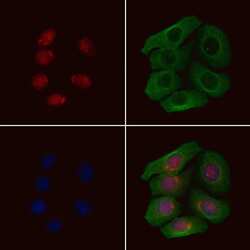
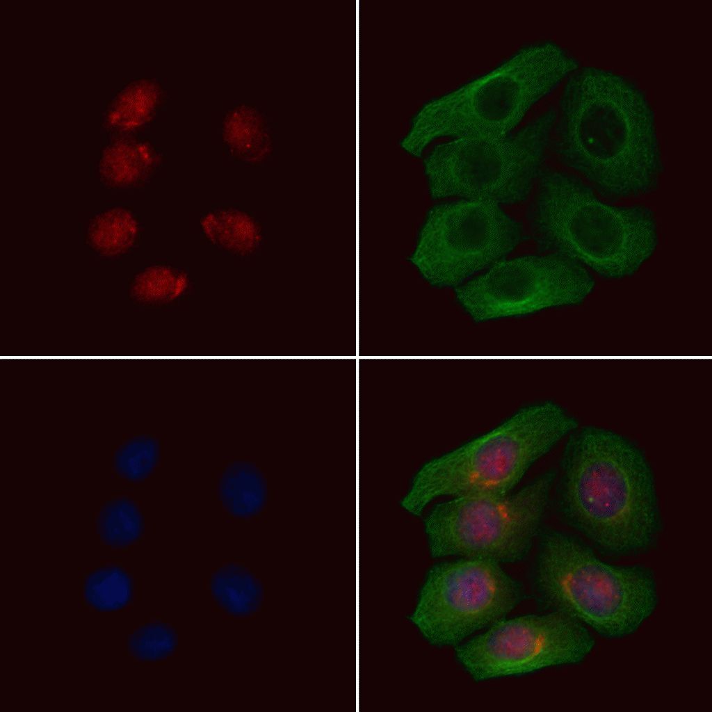
- Immunofluorescent analysis of Phospho-IRF5 (Ser437) in HeLa cells(4h of LPS treatment). Samples were fixed with paraformaldehyde, permeabilized with 0.1% Triton X-100, blocked with 10% serum (45 min at 25°C), incubated with mouse anti-beta tubulin and Phospho-IRF5 (Ser437) polyclonal antibody (Product # PA5-106093) using a dilution of 1:200 (1 hr, 37°C), and followed by goat anti-rabbit IgG Alexa Fluor 594 (red) and goat anti-mouse IgG Alexa Fluor 488 (green).
- Submitted by
- Invitrogen Antibodies (provider)
- Main image
- Experimental details
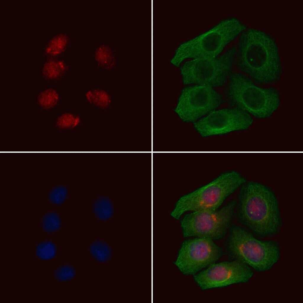
- Immunofluorescent analysis of Phospho-IRF5 (Ser437) in HeLa cells(4h of LPS treatment). Samples were fixed with paraformaldehyde, permeabilized with 0.1% Triton X-100, blocked with 10% serum (45 min at 25°C), incubated with mouse anti-beta tubulin and Phospho-IRF5 (Ser437) polyclonal antibody (Product # PA5-106093) using a dilution of 1:200 (1 hr, 37°C), and followed by goat anti-rabbit IgG Alexa Fluor 594 (red) and goat anti-mouse IgG Alexa Fluor 488 (green).
Supportive validation
- Submitted by
- Invitrogen Antibodies (provider)
- Main image
- Experimental details
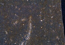
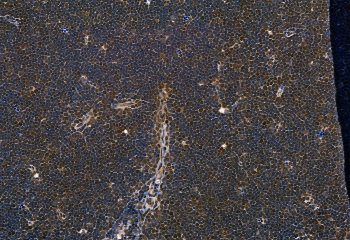
- Immunohistochemistry analysis of Phospho-IRF5 (Ser437) in rat thymus tissue. The sample was formaldehyde fixed and a heat mediated antigen retrieval step in citrate buffer was performed. Samples were incubated with Phospho-IRF5 (Ser437) polyclonal antibody (Product # PA5-106093) using a dilution of 1:100 (4°C overnight) followed by HRP conjugated anti-Rabbit secondary antibody.
Supportive validation
- Submitted by
- Invitrogen Antibodies (provider)
- Main image
- Experimental details
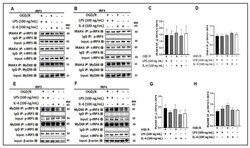
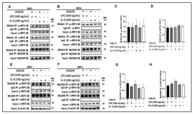
- Figure 1 IRF5 and IRF4 bind to MyD88 or IRAK4. ( A , B ) SIM-A9 cell homogenates were subjected to Co-IP with anti-IRAK4 antibody followed by immunoblotting for p-IRF5/p-IRF4, t-IRF5/t-IRF4, and MyD88. ( C , D ) WB optical density quantification of the ratio of p-IRF5 over t-IRF5 ( C ) and p-IRF4 over t-IRF4 ( D ). ( E , F ) SIM-A9 cell homogenates were subjected to Co-IP with anti-MyD88 antibody to detect p-IRF5/p-IRF4 and t-IRF5/t-IRF4. ( G , H ) WB optical density quantification of the ratio of p-IRF5 over t-IRF5 ( G ) and p-IRF4 over t-IRF4 ( H ). IgG controls were from the same homogenates of each treatment. n = 3 independent experiments/per condition. IB, immunoblot; p, phosphorylated; t, total; IgG, immunoglobulin negative control. One-way ANOVA.
- Submitted by
- Invitrogen Antibodies (provider)
- Main image

- Experimental details
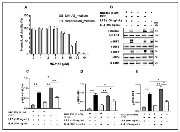
- Figure 2 IRAK4 phosphorylates IRF5/IRF4. ( A ) SIM-A9 cells were treated with 8 muM ND2158, chosen by MTS gradient viability assay. ( B ) After treatments, cell homogenates were immunoblotted. ImageJ quantified ratios of p-IRAK4/t-IRAK4 ( C ), p-IRF5/t-IRF5 ( D ), and p-IRF4/t-IRF4 ( E ) in ( B ). Data were obtained from 3 independent experiments. p, phosphorylated; t, total. ** p < 0.0001, * p < 0.05; One-way ANOVA.
- Submitted by
- Invitrogen Antibodies (provider)
- Main image
- Experimental details
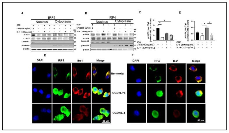
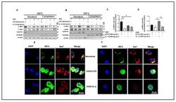
- Figure 3 Microglial IRF5 and IRF4 translocate from the cytoplasm to the nucleus. ( A, B ) Cell homogenates were fractionated to nuclear and cytoplasmic portions and Western blot performed in each fraction for p-IRF5/4 and t-IRF5/4. The OD of each band was normalized to beta-actin first, and then the quantitative ratio of nuclear/cytoplasmic p-IRF5 ( C ) and nuclear/cytoplasmic p-IRF4 ( D ) were presented. Immunocytochemistry for IRF5 ( E ) and IRF4 ( F ) in microglia showing cytosolic and nuclear expression. Western blot data were from 3 independent experiments. * p < 0.05; One-way ANOVA. Lamin b1 and beta-tubulin are for the purity validation of nuclear and cytoplasmic fraction respectively. p , phosphorylated; t , total.