 Explore
 Validate
 Learn
Learn Western blot
Antibody data
- Antibody Data
- Antigen structure
- References [0]
- Comments [0]
- Validations
- Western blot [2]
- Immunocytochemistry [1]
Submit
Validation data
Reference
Comment
Report error
- Product number
- AF4508 - Provider product page

- Provider
- R&D Systems
- Product name
- Human IRF5 Antibody
- Antibody type
- Polyclonal
- Description
- Antigen Affinity-purified. Detects human IRF5 in Western blots.
- Reactivity
- Human
- Host
- Sheep
- Conjugate
- Unconjugated
- Antigen sequence
NP_002191- Isotype
- IgG
- Vial size
- 100 ug
- Concentration
- LYOPH
- Storage
- Use a manual defrost freezer and avoid repeated freeze-thaw cycles. 12 months from date of receipt, -20 to -70 °C as supplied. 1 month, 2 to 8 °C under sterile conditions after reconstitution. 6 months, -20 to -70 °C under sterile conditions after reconstitution.
No comments: Submit comment
Supportive validation
- Submitted by
- R&D Systems (provider)
- Main image
- Experimental details
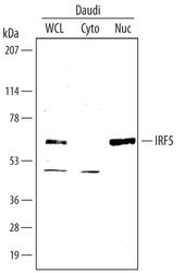
- Detection of Human IRF5 by Western Blot. Western blot shows lysates of Daudi human Burkitt's lymphoma cell line. Gels were loaded with 30 μg of whole cell lysate (WCL), 20 μg of cytoplasmic (Cyto), and 10 μg of nuclear extracts (Nuc). PVDF membrane was probed with 2 µg/mL Sheep Anti-Human IRF5 Antigen Affinity-purified Polyclonal Antibody (Catalog # AF4508) followed by HRP-conjugated Anti-Mouse IgG Secondary Antibody (Catalog # HAF007). A specific band for IRF5 was detected at approximately 58 kDa (as indicated). This experiment was conducted under reducing conditions and using Immunoblot Buffer Group 3.
- Submitted by
- R&D Systems (provider)
- Main image
- Experimental details
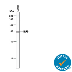
- Detection of Human IRF5 by Simple WesternTM. Simple Western lane view shows lysates of Daudi human Burkitt's lymphoma cell line, loaded at 0.2 mg/mL. A specific band was detected for IRF5 at approximately 64 kDa (as indicated) using 20 µg/mL of Sheep Anti-Human IRF5 Antigen Affinity-purified Polyclonal Antibody (Catalog # AF4508) followed by 1:50 dilution of HRP-conjugated Anti-Sheep IgG Secondary Antibody (Catalog # HAF016). This experiment was conducted under reducing conditions and using the 12-230 kDa separation system.
Supportive validation
- Submitted by
- R&D Systems (provider)
- Main image
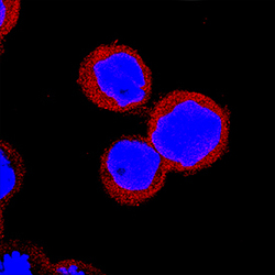
- Experimental details
- IRF5 in THP-1 Human Cell Line. IRF5 was detected in immersion fixed THP-1 human acute monocytic leukemia cell line using Sheep Anti-Human IRF5 Antigen Affinity-purified Polyclonal Antibody (Catalog # AF4508) at 15 µg/mL for 3 hours at room temperature. Cells were stained using the NorthernLights™ 557-conjugated Anti-Sheep IgG Secondary Antibody (red; Catalog # NL010) and counterstained with DAPI (blue). Specific staining was localized to cytoplasm. View our protocol for Fluorescent ICC Staining of Non-adherent Cells.
