Explore
 Validate
 Learn
Learn Western blot
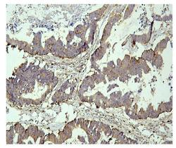
 Immunohistochemistry
Antibody data
- Antibody Data
- Antigen structure
- References [1]
- Comments [0]
- Validations
- Western blot [1]
Submit
Validation data
Reference
Comment
Report error
- Product number
- M02690 - Provider product page

- Provider
- Boster Biological Technology
- Product name
- Anti-PPT1 Antibody Picoband™ (monoclonal, 10F3)
- Antibody type
- Monoclonal
- Description
- Mouse IgG monoclonal antibody for PPT1 detection. Tested with WB, IHC-P, FCM in Human.
- Reactivity
- Human
- Host
- Mouse
- Isotype
- IgG
- Antibody clone number
- 10F3
- Vial size
- 100μg/vial
- Concentration
- 0.5-1mg/ml, actual concentration vary by lot. Use suggested dilution ratio to decide dilution procedure.
- Storage
- At -20°C for one year. After reconstitution, at 4°C for one month. It can also be aliquoted and stored frozen at -20°C for a longer time. Avoid repeated freezing and thawing.
- Handling
- Add 0.2ml of distilled water will yield a concentration of 500μg/ml.
Submitted references High PPT1 expression predicts poor clinical outcome and PPT1 inhibitor DC661 enhances sorafenib sensitivity in hepatocellular carcinoma.
Xu J, Su Z, Cheng X, Hu S, Wang W, Zou T, Zhou X, Song Z, Xia Y, Gao Y, Zheng Q
Cancer cell international 2022 Mar 11;22(1):115
Cancer cell international 2022 Mar 11;22(1):115
No comments: Submit comment
Supportive validation
- Submitted by
- Boster Biological Technology (provider)
- Main image

- Experimental details
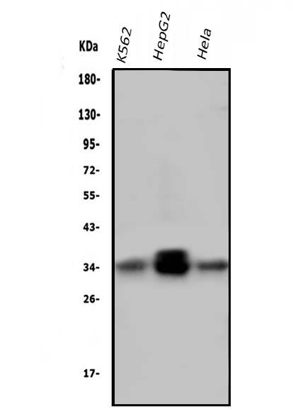
- Western blot analysis of PPT1 using anti-PPT1 antibody (M02690). Electrophoresis was performed on a 5-20% SDS-PAGE gel at 70V (Stacking gel) / 90V (Resolving gel) for 2-3 hours. The sample well of each lane was loaded with 50ug of sample under reducing conditions. Lane 1: human K562 whole cell lysates, Lane 2: human HepG2 whole cell lysates, Lane 3: human Hela whole cell lysates. After Electrophoresis, proteins were transferred to a Nitrocellulose membrane at 150mA for 50-90 minutes. Blocked the membrane with 5% Non-fat Milk/ TBS for 1.5 hour at RT. The membrane was incubated with mouse anti-PPT1 antigen affinity purified monoclonal antibody (Catalog # M02690) at 0.5 μg/mL overnight at 4°C, then washed with TBS-0.1%Tween 3 times with 5 minutes each and probed with a goat anti-mouse IgG-HRP secondary antibody at a dilution of 1:10000 for 1.5 hour at RT. The signal is developed using an Enhanced Chemiluminescent detection (ECL) kit (Catalog # EK1001) with Tanon 5200 system. A specific band was detected for PPT1 at approximately 34KD. The expected band size for PPT1 is at 34KD.
- Additional image