Explore
 Validate
 Learn
Learn Western blot
 Immunoprecipitation
Antibody data
- Antibody Data
- Antigen structure
- References [3]
- Comments [0]
- Validations
- Western blot [2]
- Immunohistochemistry [1]
- Blocking/Neutralizing [1]
Submit
Validation data
Reference
Comment
Report error
- Product number
- AF907 - Provider product page

- Provider
- R&D Systems
- Product name
- Human MMP-7 Antibody
- Antibody type
- Polyclonal
- Description
- Antigen Affinity-purified. Detects human MMP-7 in direct ELISAs and Western blots. In direct ELISAs, approximately 50% cross-reactivity with recombinant mouse MMP-7 is observed, and less than 1% cross-reactivity with recombinant human (rh) MMP-8 and rhMMP-12 is observed.
- Reactivity
- Human
- Host
- Goat
- Conjugate
- Unconjugated
- Antigen sequence
P09237- Isotype
- IgG
- Vial size
- 100 ug
- Concentration
- LYOPH
- Storage
- Use a manual defrost freezer and avoid repeated freeze-thaw cycles. 12 months from date of receipt, -20 to -70 °C as supplied. 1 month, 2 to 8 °C under sterile conditions after reconstitution. 6 months, -20 to -70 °C under sterile conditions after reconstitution.
Submitted references Farnesoid X receptor represses matrix metalloproteinase 7 expression, revealing this regulatory axis as a promising therapeutic target in colon cancer.
Gelsolin induces colorectal tumor cell invasion via modulation of the urokinase-type plasminogen activator cascade.
Determination of matrilysin activity in gastrointestinal neoplasia.
Peng Z, Chen J, Drachenberg CB, Raufman JP, Xie G
The Journal of biological chemistry 2019 May 24;294(21):8529-8542
The Journal of biological chemistry 2019 May 24;294(21):8529-8542
Gelsolin induces colorectal tumor cell invasion via modulation of the urokinase-type plasminogen activator cascade.
Zhuo J, Tan EH, Yan B, Tochhawng L, Jayapal M, Koh S, Tay HK, Maciver SK, Hooi SC, Salto-Tellez M, Kumar AP, Goh YC, Lim YC, Yap CT
PloS one 2012;7(8):e43594
PloS one 2012;7(8):e43594
Determination of matrilysin activity in gastrointestinal neoplasia.
Hawinkels LJ, Verspaget HW, van den Berg M, Hanemaaijer R, Sier CF
European journal of clinical investigation 2007 Jul;37(7):598-9
European journal of clinical investigation 2007 Jul;37(7):598-9
No comments: Submit comment
Supportive validation
- Submitted by
- R&D Systems (provider)
- Main image
- Experimental details
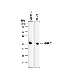
- Detection of Human MMP-7 by Western Blot. Western blot shows lysates of Capan-1 human pancreatic adenocarcinoma cell line and HT-29 human colon adenocarcinoma cell line. PVDF membrane was probed with 1 µg/mL of Goat Anti-Human MMP-7 Antigen Affinity-purified Polyclonal Antibody (Catalog # AF907) followed by HRP-conjugated Anti-Goat IgG Secondary Antibody (Catalog # HAF017). A specific band was detected for MMP-7 at approximately 28 kDa (as indicated). This experiment was conducted under reducing conditions and using Immunoblot Buffer Group 1.

- Submitted by
- R&D Systems (provider)
- Main image
- Experimental details
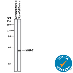
- Detection of Human MMP-7 by Simple WesternTM. Simple Western lane view shows lysates of normal stem cells (negative control) and Crohn's stem cells, loaded at 0.2 mg/mL. A specific band was detected for MMP-7 at approximately 10 kDa (as indicated) using 10 µg/mL of Goat Anti-Human MMP-7 Antigen Affinity-purified Polyclonal Antibody (Catalog # AF907) followed by 1:50 dilution of HRP-conjugated Anti-Goat IgG Secondary Antibody (Catalog # HAF109). This experiment was conducted under reducing conditions and using the 12-230 kDa separation system.

Supportive validation
- Submitted by
- R&D Systems (provider)
- Main image
- Experimental details
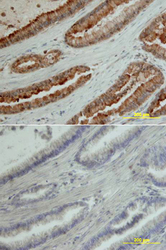
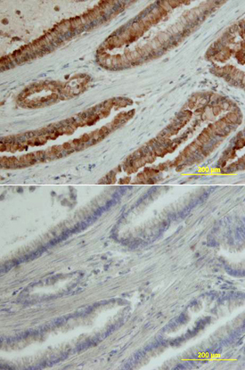
- MMP-7 in Human Pancreas. MMP-7 was detected in immersion fixed paraffin-embedded sections of human pancreas array using Goat Anti-Human MMP-7 Antigen Affinity-purified Polyclonal Antibody (Catalog # AF907) at 10 µg/mL overnight at 4 °C. Tissue was stained using the Anti-Goat HRP-DAB Cell & Tissue Staining Kit (brown; Catalog # CTS008) and counterstained with hematoxylin (blue). Lower panel shows a lack of labeling if primary antibodies are omitted and tissue is stained only with secondary antibody followed by incubation with detection reagents. View our protocol for Chromogenic IHC Staining of Paraffin-embedded Tissue Sections.
Supportive validation
- Submitted by
- R&D Systems (provider)
- Main image
- Experimental details
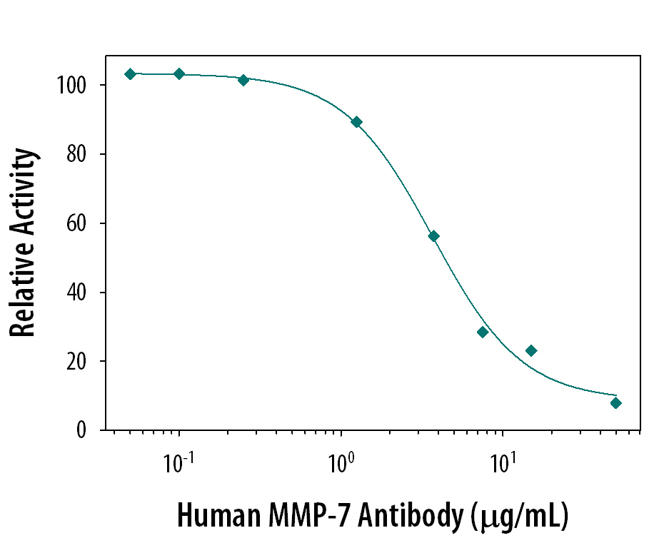
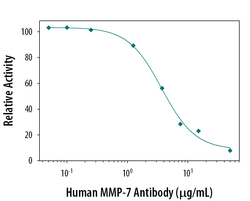
- Neutralization of MMP-7 Activity by Human MMP-7 Antibody. The cleavage of Mca-PLGL-Dpa-AR-NH2 (10 μM, Catalog # ES001) by Recombinant Human MMP-7 (0.2 µg/mL, Catalog # 907-MP) is measured after preincubation with increasing concentrations of Goat Anti-Human MMP-7 Antigen Affinity-purified Polyclonal Antibody (Catalog # AF907). The ND50 is typically 2 µg/mL.