Explore
 Validate
 Learn
Learn Immunocytochemistry
Antibody data
- Antibody Data
- Antigen structure
- References [1]
- Comments [0]
- Validations
- Immunocytochemistry [3]
- Immunohistochemistry [1]
- Other assay [1]
Submit
Validation data
Reference
Comment
Report error
- Product number
- PA5-52821 - Provider product page

- Provider
- Invitrogen Antibodies
- Product name
- SAP18 Polyclonal Antibody
- Antibody type
- Polyclonal
- Antigen
- Recombinant protein fragment
- Description
- Immunogen sequence: VTQEEIKKEP EKPIDREKTC PLLLRVFTTN NGRHHRMDEF SRGNVPSSEL QIYTWMDATL KELTSLVKEV YPEARKKGTH FNFAIVFTDV KRPGYRVKEI GSTMSGRKGT DDSMTLQSQK FQIGDYLDIA ITPPNRAPP Highest antigen sequence identity to the following orthologs: Mouse - 99%, Rat - 99%.
- Reactivity
- Human
- Host
- Rabbit
- Isotype
- IgG
- Vial size
- 100 μL
- Concentration
- 0.05 mg/mL
- Storage
- Store at 4°C short term. For long term storage, store at -20°C, avoiding freeze/thaw cycles.
Submitted references Tumor-Infiltrated CD8+ T Cell 10-Gene Signature Related to Clear Cell Renal Cell Carcinoma Prognosis.
Wang J, Huang F, Zhao J, Huang P, Tan J, Huang M, Ma R, Xiao Y, He S, Wang Z, Shen J, Lu H, Meng L
Frontiers in immunology 2022;13:930921
Frontiers in immunology 2022;13:930921
No comments: Submit comment
Supportive validation
- Submitted by
- Invitrogen Antibodies (provider)
- Main image
- Experimental details
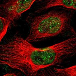
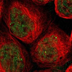
- Immunofluorescent staining of SAP18 in human cell line U-2 OS shows positivity in nucleus but excluded from the nucleoli. Samples were probed using a SAP18 Polyclonal Antibody (Product # PA5-52821).
- Submitted by
- Invitrogen Antibodies (provider)
- Main image

- Experimental details
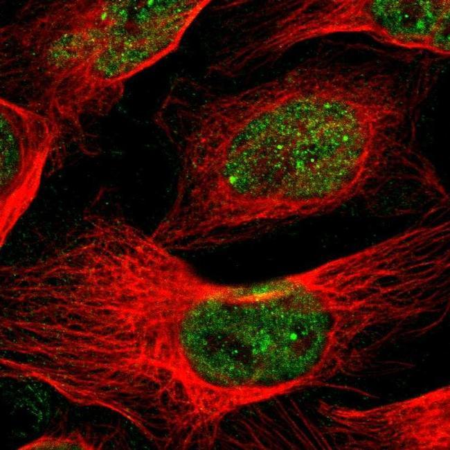
- Immunofluorescent staining of SAP18 in human cell line A-431 using a SAP18 Polyclonal Antibody (Product # PA5-52821) shows localization to nucleoplasm and nuclear bodies.
- Submitted by
- Invitrogen Antibodies (provider)
- Main image
- Experimental details
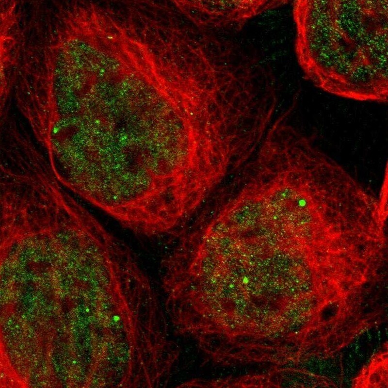
- Immunofluorescent staining of SAP18 in human cell line A-431 using a SAP18 Polyclonal Antibody (Product # PA5-52821) shows localization to nucleoplasm and nuclear bodies.

Supportive validation
- Submitted by
- Invitrogen Antibodies (provider)
- Main image
- Experimental details
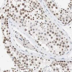
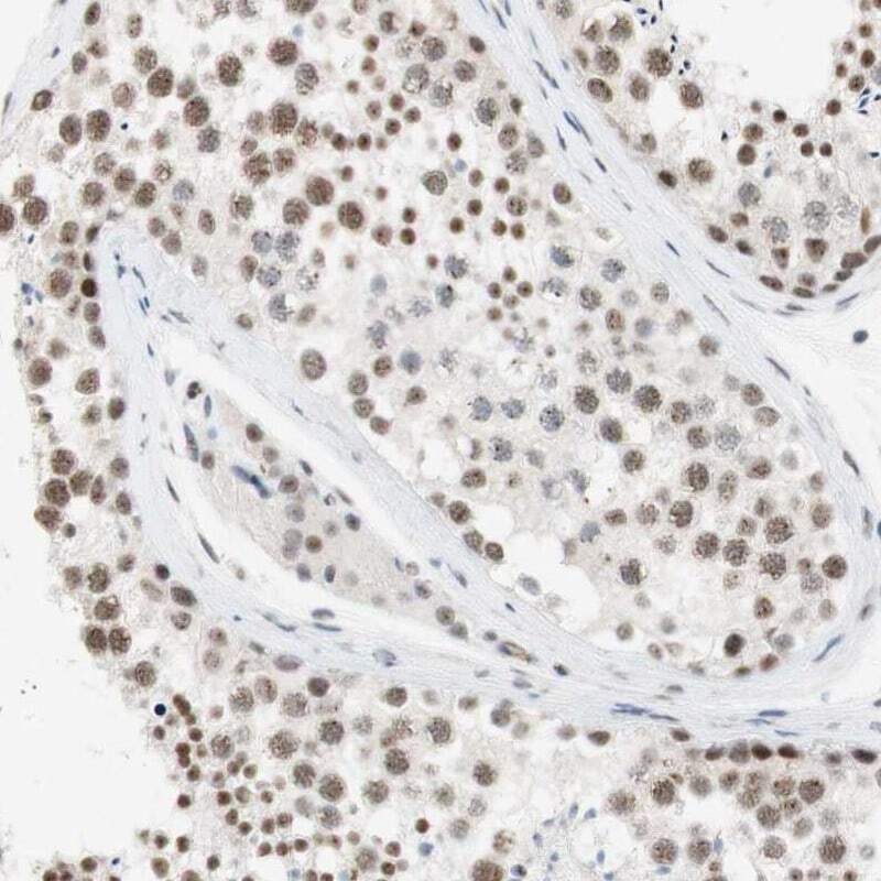
- Immunohistochemical staining of SAP18 in human testis using a SAP18 Polyclonal Antibody (Product # PA5-52821) shows nuclear positivity in cells in seminiferous ducts and Leydig cells.
Supportive validation
- Submitted by
- Invitrogen Antibodies (provider)
- Main image
- Experimental details
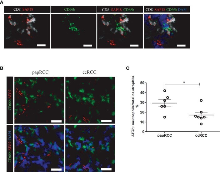
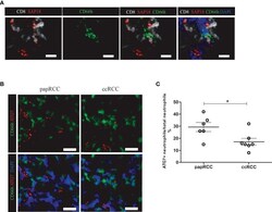
- Figure 6 SAP18-expressing CD8+ T cells co-localize with neutrophil in ccRCC biopsies. (A) Immunofluorescent imaging showing CD8+ T cells in ccRCC biopsies expressing SAP18, and co-localize with neutrophils. CD8+ T cells showing in white, SAP showing in red, neutrophils were labeled with CD66b (green), cell nucleus were counterstained with DAPI. White scale bar indicates 25 mum. (B) Immunofluorescent imaging Neutrophils (CD66b+ cell, green) in ccRCC expressing less ATG7 (red). cell nucleus were counterstained with DAPI. White scale bar indicates 25 mum. (C) Statistical analysis of the frequency of ATG7+ expressing neutrophils against total neutrophils in RCC biopsies. Each dot represented one readout. *: p