 Explore
 Validate
 Learn
Learn Western blot
 Immunocytochemistry
Antibody data
- Antibody Data
- Antigen structure
- References [3]
- Comments [0]
- Validations
- Immunocytochemistry [2]
- Immunohistochemistry [2]
- Flow cytometry [1]
- Other assay [3]
Submit
Validation data
Reference
Comment
Report error
- Product number
- 701287 - Provider product page

- Provider
- Invitrogen Antibodies
- Product name
- MMP13 Recombinant Rabbit Monoclonal Antibody (3H13L17)
- Antibody type
- Monoclonal
- Antigen
- Recombinant full-length protein
- Description
- This antibody is predicted to react with mouse, rat, non-human primate and rabbit based on sequence homology. Intact IgG appears on a non-reducing gel as ~150 kDa band and upon reduction generating a ~25 kDa light chain band and a ~50 kDa heavy chain. Recombinant rabbit monoclonal antibodies are produced using in vitro expression systems. The expression systems are developed by cloning in the specific antibody DNA sequences from immunoreactive rabbits. Then, individual clones are screened to select the best candidates for production. The advantages of using recombinant rabbit monoclonal antibodies include: better specificity and sensitivity, lot-to-lot consistency, animal origin-free formulations, and broader immunoreactivity to diverse targets due to larger rabbit immune repertoire.
- Reactivity
- Human
- Host
- Rabbit
- Isotype
- IgG
- Antibody clone number
- 3H13L17
- Vial size
- 100 μg
- Concentration
- 0.5 mg/mL
- Storage
- Store at 4°C short term. For long term storage, store at -20°C, avoiding freeze/thaw cycles.
Submitted references Xanthohumol Attenuated Inflammation and ECM Degradation by Mediating HO-1/C/EBPβ Pathway in Osteoarthritis Chondrocytes.
GYY4137 Regulates Extracellular Matrix Turnover in the Diabetic Kidney by Modulating Retinoid X Receptor Signaling.
Distinct biological events generated by ECM proteolysis by two homologous collagenases.
Zhang M, Zhang R, Zheng T, Chen Z, Ji G, Peng F, Wang W
Frontiers in pharmacology 2021;12:680585
Frontiers in pharmacology 2021;12:680585
GYY4137 Regulates Extracellular Matrix Turnover in the Diabetic Kidney by Modulating Retinoid X Receptor Signaling.
Juin SK, Pushpakumar S, Sen U
Biomolecules 2021 Oct 7;11(10)
Biomolecules 2021 Oct 7;11(10)
Distinct biological events generated by ECM proteolysis by two homologous collagenases.
Solomonov I, Zehorai E, Talmi-Frank D, Wolf SG, Shainskaya A, Zhuravlev A, Kartvelishvily E, Visse R, Levin Y, Kampf N, Jaitin DA, David E, Amit I, Nagase H, Sagi I
Proceedings of the National Academy of Sciences of the United States of America 2016 Sep 27;113(39):10884-9
Proceedings of the National Academy of Sciences of the United States of America 2016 Sep 27;113(39):10884-9
No comments: Submit comment
Supportive validation
- Submitted by
- Invitrogen Antibodies (provider)
- Main image
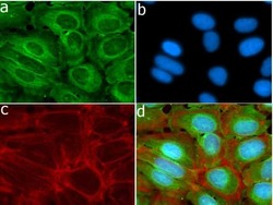
- Experimental details
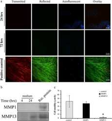
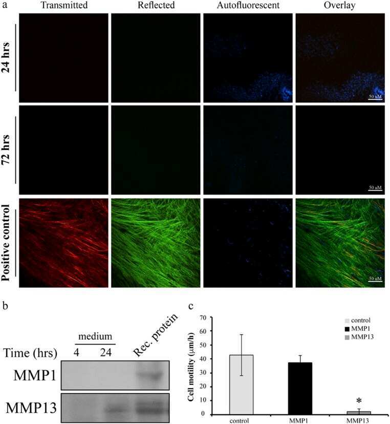
- Immunofluorescent analysis of MMP-13 in HeLa cells using a MMP-13 recombinant rabbit monoclonal antibody (Product # 701287) followed by detection using an Alexa Fluor 488-conjugated goat anti-rabbit secondary antibody (green) (Image A). Nuclei were stained using DAPI (Image B) and actin stained with Alexa Fluor 594 phalloidin (red) (image C). Image D is a composite image showing nuclear localization of MMP-13.

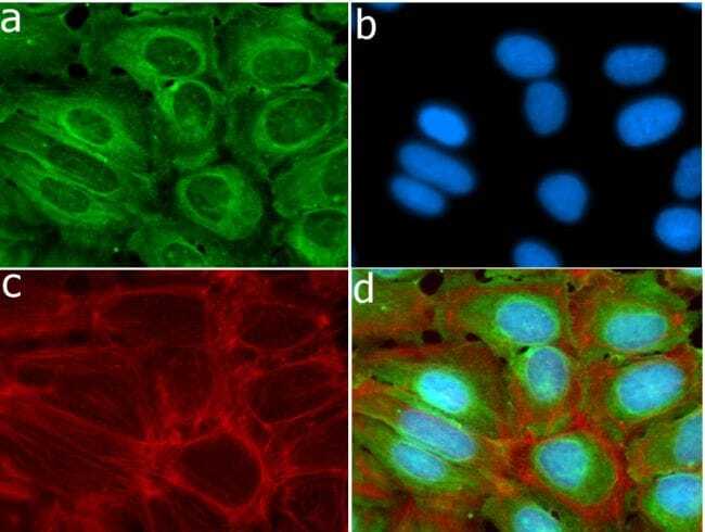
- Submitted by
- Invitrogen Antibodies (provider)
- Main image
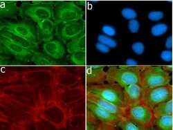
- Experimental details
- Immunofluorescent analysis of MMP-13 in HeLa cells using a MMP-13 recombinant rabbit monoclonal antibody (Product # 701287) followed by detection using an Alexa Fluor 488-conjugated goat anti-rabbit secondary antibody (green) (Image A). Nuclei were stained using DAPI (Image B) and actin stained with Alexa Fluor 594 phalloidin (red) (image C). Image D is a composite image showing nuclear localization of MMP-13.
Supportive validation
- Submitted by
- Invitrogen Antibodies (provider)
- Main image
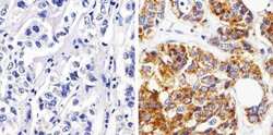
- Experimental details
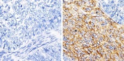
- Immunohistochemistry analysis of MMP-13/Collagenase-3 showing staining in the cytoplasm of paraffin-embedded human breast carcinoma (right) compared to a negative control without primary antibody (left). To expose target proteins, antigen retrieval was performed using 10 mM sodium citrate (pH 6.0), microwaved for 8-15 min. Following antigen retrieval, tissues were blocked in 3% H2O2-methanol for 15 min at room temperature, washed with ddH2O and PBS, and then probed with MMP-13/Collagenase-3 Monoclonal antibody (Product # 701287) diluted in 3% BSA-PBS at a dilution of 1:50 overnight at 4°C in a humidified chamber. Tissues were washed extensively in PBST and detection was performed using a HRP-conjugated secondary antibody followed by colorimetric detection using a DAB kit. Tissues were counterstained with hematoxylin and dehydrated with ethanol and xylene to prep for mounting.

- Submitted by
- Invitrogen Antibodies (provider)
- Main image
- Experimental details
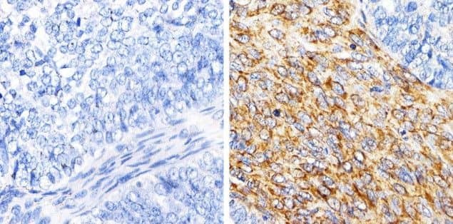
- Immunohistochemistry analysis of MMP-13/Collagenase-4 showing staining in the cytoplasm of paraffin-embedded human ovarian carcinoma (right) compared to a negative control without primary antibody (left). To expose target proteins, antigen retrieval was performed using 10 mM sodium citrate (pH 6.0), microwaved for 8-15 min. Following antigen retrieval, tissues were blocked in 3% H2O2-methanol for 15 min at room temperature, washed with ddH2O and PBS, and then probed with MMP-13/Collagenase-4 monoclonal antibody (Product # 701287) diluted in 3% BSA-PBS at a dilution of 1:100 overnight at 4°C in a humidified chamber. Tissues were washed extensively in PBST and detection was performed using a HRP-conjugated secondary antibody followed by colorimetric detection using a DAB kit. Tissues were counterstained with hematoxylin and dehydrated with ethanol and xylene to prep for mounting.
Supportive validation
- Submitted by
- Invitrogen Antibodies (provider)
- Main image
- Experimental details
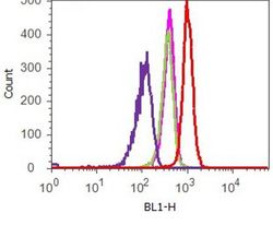
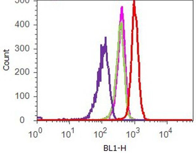
- Flow cytometry analysis of MMP13 was done on HeLa cells. Cells were fixed with 70% ethanol for 10 minutes, permeabilized with 0.25% Tritonª X-100 for 20 minutes, and blocked with 5% BSA for 1 hour at room temperature. Cells were labeled with ABfinityª MMP13 Recombinant Rabbit Monoclonal Antibody (701287, red histogram) or with rabbit isotype control (pink histogram) at 2 µg-4 µg/million cells in 2.5% BSA. After incubation at room temperature for 2-3 hours, the cells were labeled with Alexa Fluor¨ 488 Goat Anti-Rabbit Secondary Antibody (A11008) at a dilution of 1:400 for 30 minutes at room temperature. The representative 10,000 cells were acquired and analyzed for each sample using an Attune¨ Acoustic Focusing Cytometer. The purple histogram represents unstained control cells and the green histogram represents no-primary-antibody control.
Supportive validation
- Submitted by
- Invitrogen Antibodies (provider)
- Main image
- Experimental details
- NULL
- Submitted by
- Invitrogen Antibodies (provider)
- Main image
- Experimental details
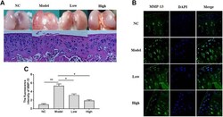
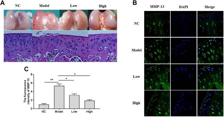
- FIGURE 1 The gross observation, histomorphological examination, and immunofluorescence assays in the knee articular cartilage. (A) The gross observation and histomorphological examination (HE staining) of articular cartilage. (B) The immunofluorescence study of MMP-13 in cartilage. (C) The summary data of fluorescence intensity of MMP-13 in situ . All experiments were performed in triplicate and data are presented as the mean +- standard deviation. * p < 0.05 and ** p < 0.01. NC, negative control; Model, the model group; Low, the group treated with XH (5.64 mg/kg); High, the group treated with XH (16.9 mg/kg).
- Submitted by
- Invitrogen Antibodies (provider)
- Main image
- Experimental details
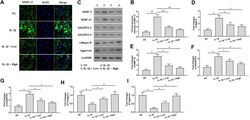
- FIGURE 4 XH attenuated ECM degradation in IL-1beta-treated chondrocytes. (A) The immunofluorescence study of MMP-13 in IL-1beta-treated chondrocytes. (B) The summary data of fluorescence intensity of MMP-13. (C) The proteins expression of MMP-3, MMP-13, ADAMTS-4, ADAMTS-5, collagen-II, and aggrecan were detected by western blotting. (D-I) were the quantified values of tested proteins. All experiments were performed in triplicate and data are presented as the mean +- standard deviation. * p < 0.05 and ** p < 0.01. NC, negative control; Low, XH (5 muM); High, XH (20 muM).
