Explore
 Validate
 Learn
Learn Western blot
Antibody data
- Antibody Data
- Antigen structure
- References [1]
- Comments [0]
- Validations
- Western blot [2]
- Immunocytochemistry [1]
- Other assay [1]
Submit
Validation data
Reference
Comment
Report error
- Product number
- PA5-16566 - Provider product page

- Provider
- Invitrogen Antibodies
- Product name
- MMP13 Polyclonal Antibody
- Antibody type
- Polyclonal
- Antigen
- Recombinant full-length protein
- Description
- PA5-16566 targets MMP-13 (Collagenase-3) in WB applications and shows reactivity with Human and Rat samples.
- Concentration
- 1 mg/mL
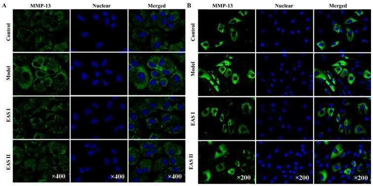
Submitted references Electroacupuncture serum inhibits TNF‑α‑mediated chondrocyte inflammation via the Ras‑Raf‑MEK1/2‑ERK1/2 signaling pathway.
Chen H, Shao X, Li L, Zheng C, Xu X, Hong X, Li X, Wu M
Molecular medicine reports 2017 Nov;16(5):5807-5814
Molecular medicine reports 2017 Nov;16(5):5807-5814
No comments: Submit comment
Supportive validation
- Submitted by
- Invitrogen Antibodies (provider)
- Main image
- Experimental details
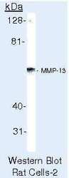
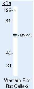
- Western blot of MMP-13 (Collagenase-3) using MMP-13 (Collagenase-3) Polyclonal Antibody (Product # PA5-16566) on CCL-4 med Cells.
- Submitted by
- Invitrogen Antibodies (provider)
- Main image

- Experimental details
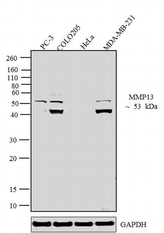
- Western blot analysis was performed on whole cell extracts (30 µg lysate) of PC-3 (Lane 1), COLO205 (Lane 2), HeLa (Lane 3) and MDA-MB-231 (Lane4). The blots were probed with Anti MMP-13 Rabbit Polyclonal Antibody (Product # PA5-16566, 1-3 µg/mL) and detected by chemiluminescence using Goat anti-Rabbit IgG (H+L) Secondary Antibody, HRP conjugate (Product # G-21234, 1:5000 dilution). A 53 kDa band corresponding to MMP-13 was observed across cell lines tested except HeLa. An active form of the protein ~40 kDa was seen in COLO205 and MDA-MB-231. Known quantity of protein samples were electrophoresed using Novex® NuPAGE® 10 % Bis-Tris gel (Product # NP0301BOX), XCell SureLock™ Electrophoresis System (Product # EI0002) and Novex® Sharp Pre-Stained Protein Standard (Product # LC5800). Resolved proteins were then transferred onto a nitrocellulose membrane with PierceTM Power Blotter System (Product # 22834). The membrane was probed with the relevant primary and secondary Antibody following blocking with 5 % skimmed milk. Chemiluminescent detection was performed using Pierce™ ECL Western Blotting Substrate (Product # 32106).
Supportive validation
- Submitted by
- Invitrogen Antibodies (provider)
- Main image
- Experimental details
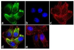
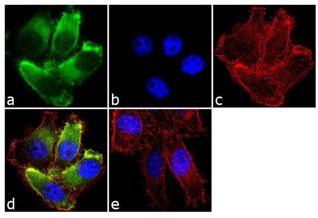
- Immunofluorescence analysis of MMP-13 (Collagenase-3) was done on 70% confluent log phase PC-3 cells. The cells were fixed with 4% paraformaldehyde for 10 minutes, permeabilized with 0.1% Triton™ X-100 for 10 minutes, and blocked with 1% BSA for 1 hour at room temperature. The cells were labeled with MMP-13 (Collagenase-3) Rabbit Polyclonal Antibody (Product # PA5-16566) at 2 µg/mL in 0.1% BSA and incubated for 3 hours at room temperature and then labeled with Goat anti-Rabbit IgG (H+L) Superclonal™ Secondary Antibody, Alexa Fluor® 488 conjugate (Product # A27034) at a dilution of 1:2000 for 45 minutes at room temperature (Panel a: green). Nuclei (Panel b: blue) were stained with SlowFade® Gold Antifade Mountant with DAPI (Product # S36938). F-actin (Panel c: red) was stained with Alexa Fluor® 555 Rhodamine Phalloidin (Product # R415, 1:300). Panel d is a merged image showing cytoplasmic localization. Panel e is a no primary antibody control. The images were captured at 60X magnification.
Supportive validation
- Submitted by
- Invitrogen Antibodies (provider)
- Main image
- Experimental details
- NULL