Explore
 Validate
 Learn
Learn Western blot
Antibody data
- Antibody Data
- Antigen structure
- References [0]
- Comments [0]
- Validations
- Western blot [1]
Submit
Validation data
Reference
Comment
Report error
- Product number
- 200-401-913 - Provider product page

- Provider
- Rockland Immunochemicals, Inc.
- Proper citation
- Rockland Cat#200-401-913, RRID:AB_2182938
- Product name
- Anti-SUMO Activating Enzyme E1 (RABBIT) Antibody - 200-401-913
- Antibody type
- Polyclonal
- Vial size
- 500 µl
No comments: Submit comment
Supportive validation
- Submitted by
- Rockland Immunochemicals, Inc. (provider)
- Main image

- Experimental details
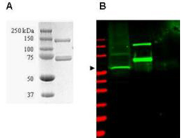
- Coommassie-stained SDS-PAGE of GST-SAE1 recombinant protein (Panel A) and western blotting (Panel B) of HeLa WC lysate (lane 1) and purified recombinant GST-SAE1 (lane 2) are presented to show specificity of Rockland's purified anti-SUMO Activating Enzyme (SAE1) antibody. The recombinant protein (with tag) ~60 kDa band present in 35 µg lysate (green, 800 nm channel) is indicated by the arrowhead. Lane 2 contains 50 ng of purified recombinant GST-SAE1 and lane 3 contains 300 ng of purified GST. Proteins were separated on a 4-20% Tris-Glycine gel by SDS-PAGE and transferred onto nitrocellulose. After blocking the membrane was probed with the primary antibody diluted to 1:2,000. Incubation was overnight at 4° C followed by washes and reaction with a 1:10,000 dilution of IRDye800 conjugated Gt-a-Rabbit IgG [H&L] MXHu (611-432-122) for 45 min at room temperature. Molecular weight markers are shown for both the Coommassie-stained gel and the western blot (lane M, red, 700 nm channel). IRDye800 fluorescence image was captured using the Odyssey® Infrared Imaging System developed by LI-COR. IRDye is a trademark of LI-COR, Inc. Other detection systems will yield similar results. SDS-PAGE image courtesy of Proteome Resources, Englewood, CO, http://www.proteomeresources.com.
- Validation comment
- Western Blot