Explore
 Validate
 Learn
Learn Western blot
Antibody data
- Antibody Data
- Antigen structure
- References [7]
- Comments [0]
- Validations
- Western blot [4]
- Immunocytochemistry [1]
- Other assay [5]
Submit
Validation data
Reference
Comment
Report error
- Product number
- 700791 - Provider product page

- Provider
- Invitrogen Antibodies
- Product name
- Securin Recombinant Rabbit Monoclonal Antibody (19H16L48)
- Antibody type
- Monoclonal
- Antigen
- Recombinant full-length protein
- Description
- This antibody is predicted to react with bovine based on sequence homology.
- Antibody clone number
- 19H16L48
- Concentration
- 0.5 mg/mL
Submitted references Accumulation of Securin on Spindle During Female Meiosis I.
Age-dependent integrity of the meiotic spindle assembly checkpoint in females requires Aurora kinase B.
A unique binding mode of Nek2A to the APC/C allows its ubiquitination during prometaphase.
Cyclin A2 degradation during the spindle assembly checkpoint requires multiple binding modes to the APC/C.
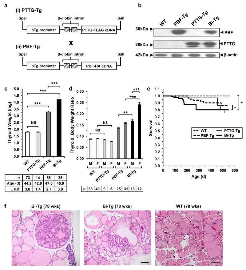
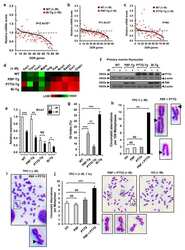
Elevated PTTG and PBF predicts poor patient outcome and modulates DNA damage response genes in thyroid cancer.
Molecular basis of APC/C regulation by the spindle assembly checkpoint.
Potential new chemotherapy strategy for human ovarian carcinoma with a novel KSP inhibitor.
Pauerova T, Radonova L, Horakova A, Knott JG, Anger M
Frontiers in cell and developmental biology 2021;9:701179
Frontiers in cell and developmental biology 2021;9:701179
Age-dependent integrity of the meiotic spindle assembly checkpoint in females requires Aurora kinase B.
Blengini CS, Nguyen AL, Aboelenain M, Schindler K
Aging cell 2021 Nov;20(11):e13489
Aging cell 2021 Nov;20(11):e13489
A unique binding mode of Nek2A to the APC/C allows its ubiquitination during prometaphase.
Alfieri C, Tischer T, Barford D
EMBO reports 2020 Jun 4;21(6):e49831
EMBO reports 2020 Jun 4;21(6):e49831
Cyclin A2 degradation during the spindle assembly checkpoint requires multiple binding modes to the APC/C.
Zhang S, Tischer T, Barford D
Nature communications 2019 Aug 27;10(1):3863
Nature communications 2019 Aug 27;10(1):3863
Elevated PTTG and PBF predicts poor patient outcome and modulates DNA damage response genes in thyroid cancer.
Read ML, Fong JC, Modasia B, Fletcher A, Imruetaicharoenchoke W, Thompson RJ, Nieto H, Reynolds JJ, Bacon A, Mallick U, Hackshaw A, Watkinson JC, Boelaert K, Turnell AS, Smith VE, McCabe CJ
Oncogene 2017 Sep 14;36(37):5296-5308
Oncogene 2017 Sep 14;36(37):5296-5308
Molecular basis of APC/C regulation by the spindle assembly checkpoint.
Alfieri C, Chang L, Zhang Z, Yang J, Maslen S, Skehel M, Barford D
Nature 2016 Aug 25;536(7617):431-436
Nature 2016 Aug 25;536(7617):431-436
Potential new chemotherapy strategy for human ovarian carcinoma with a novel KSP inhibitor.
Takenaga M, Yamamoto Y, Takeuchi T, Ohta Y, Tokura Y, Hamaguchi A, Asai D, Nakashima H, Oishi S, Fujii N
Biochemical and biophysical research communications 2015 Jul 31;463(3):222-8
Biochemical and biophysical research communications 2015 Jul 31;463(3):222-8
No comments: Submit comment
Supportive validation
- Submitted by
- Invitrogen Antibodies (provider)
- Main image
- Experimental details
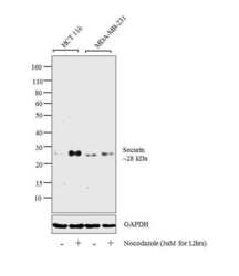
- Western blot analysis was performed on whole cell extracts (30 µg lysate) of HCT 116 (Lane 1), HCT 116 treated with Nocodazole (3uM for 12 hrs) (Lane 2), MDA-MB-231 (Lane 3) and MDA-MB-231 treated with Nocodazole (3uM for 12 hrs) (Lane 4). The blot was probed with Anti-Securin Recombinant Rabbit Monoclonal Antibody (Product # 700791, 1µg/ml) and detected by chemiluminescence using Goat anti-Rabbit IgG (H+L) Superclonal™ Secondary Antibody, HRP conjugate (Product # A27036, 0.25 µg/ml, 1:4000 dilution). An induction in 28 kDa band corresponding to Securin was observed across the cell lines tested upon treatment with nocodazole.

- Submitted by
- Invitrogen Antibodies (provider)
- Main image
- Experimental details
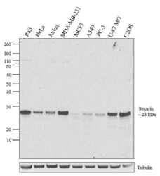
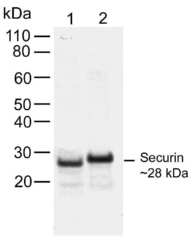
- Western blot analysis of Securin was performed by loading 20 µg of Raji (lane1), HeLa (lane2), Jurkat (lane3), MDA-MB-231 (lane4), MCF7 (lane5), A549 (lane6), PC-3 (lane7), U-87 MG (lane8) and U2OS (lane9) cell lysate using Novex® NuPAGE® 4-12 % Bis-Tris gel (Product # NP0321BOX), XCell SureLock Electrophoresis System (Product # EI0002), Novex® Sharp Pre-Stained Protein Standard (LC5800), and iBlot® Dry Blotting System (IB21001). Proteins were transferred to a nitrocellulose membrane and blocked with 5 % skim milk for 1 hour at room temperature. Securin was detected at ~28 kDa using Securin Mouse monoclonal Antibody (Product # 700791) at 1-3 µg/mL in 2.5 % skim milk at 4°C overnight on a rocking platform. Goat Anti-Rabbit IgG-HRP Secondary Antibody (G21234) at 1:5000 dilution was used and chemiluminescent detection was performed using Pierce™ ECL Western Blotting Substrate (Product # 32106).
- Submitted by
- Invitrogen Antibodies (provider)
- Main image
- Experimental details
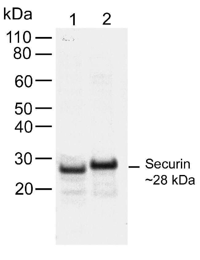
- Western blot analysis of Securin in Jurkat cell lysate (lane 1) and mouse testis lysate (lane 2) using a Securin recombinant rabbit monoclonal antibody (Product # 700791) at a dilution of 1 µg/mL. NBT/BCIP was used as the substrate (Product # WB7105).
- Submitted by
- Invitrogen Antibodies (provider)
- Main image

- Experimental details
- Knockdown of Securin was achieved by transfecting HeLa cells with Securin specific siRNAs (Silencer® select Product # s17653). Western blot analysis (Fig. a) was performed using whole cell extracts from the Securin knockdown cells (lane 3), non-specific scrambled siRNA transfected cells (lane 2) and untransfected cells (lane 1). The blot was probed with Securin Recombinant Rabbit Monoclonal Antibody (Product # 700791, 1µg/ml) and Goat anti-Rabbit IgG (H+L) Superclonal™ Secondary Antibody, HRP conjugate (Product # A27036, 0.25µg/ml, 1:4000 dilution). Densitometric analysis of this Western blot is shown in histogram (Fig. b). Decrease in signal upon siRNA mediated knock down confirms that antibody is specific to Securin.

Supportive validation
- Submitted by
- Invitrogen Antibodies (provider)
- Main image

- Experimental details
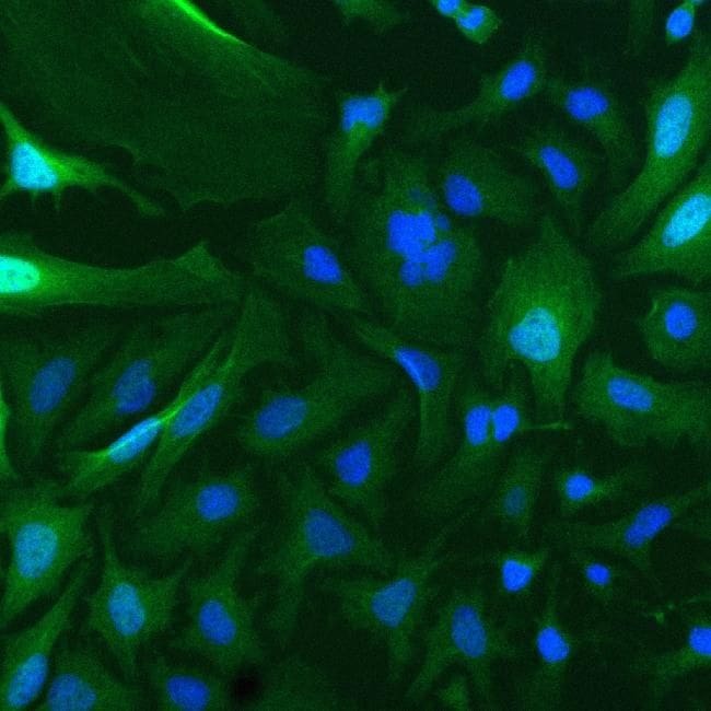
- Immunofluorescent analysis of Securin in HeLa cells using a Securin recombinant rabbit monoclonal antibody (Product # 700791) at a dilution of 2 µg/mL followed by detection using an Alexa Fluor 488-conjugated goat anti-rabbit secondary antibody at a dilution of 1:1000. Cells were fixed using 4% paraformaldehyde. The cytoplasmic localization of Securin is shown in green, while nuclei were stained using SlowFade GOLD with DAPI (Product # S36938), shown in blue.
Supportive validation
- Submitted by
- Invitrogen Antibodies (provider)
- Main image

- Experimental details
- NULL
- Submitted by
- Invitrogen Antibodies (provider)
- Main image
- Experimental details
- NULL
- Submitted by
- Invitrogen Antibodies (provider)
- Main image
- Experimental details
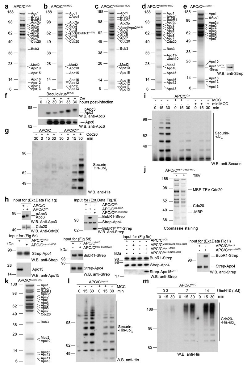
- Extended Data Figure 1 Biochemical characterization of recombinant APC/C MCC complex and preparations of wild type and mutant complexes. a,b,c,d,e SDS PAGE gels (stained with coomassie) of gel filtration peak fraction from wild type and mutant APC/C MCC complexes preparations used in this study. Western blot against Strep tag in (e) confirms the presence of Apc15 DeltaNTH-Strep .construct. f , Top: Western blot performed with anti-Apc3 antibody to monitor the time-dependent phosphorylation of this subunit induced by okadaic acid (OA) treatment in the APC/C-expressing insect cells. Bottom: Western blot against was used as a loading control and reflects the decrease in cell viability after addition of OA (data not shown). g , Western blot against His 6 -tagged ubiquitin of in vitro securin ubiquitination assays performed with either APC/C or APC/C OA in either the presence or absence of Cdc20. h , the input sample for the ubiquitination assays performed in this study is shown. i , Western blot against securin of in vitro securin ubiquitination assays performed with APC/C OA and Cdc20 with or without either MCC or miniMCC. j , SDS PAGE of APC/C MCC reconstituted with MBP-TEV-Cdc20 APC/C and untagged Cdc20 MCC . MBP-TEV-Cdc20 APC/C TEV cleavage products are indicated. k , SDS PAGE gels of reconstituted APC/C DeltaApc15-MCC complex. l , Western blot against His 6 -tagged ubiquitin of in vitro securin ubiquitination assays performed with APC/C DeltaApc15 and Cdc20 with or without M
- Submitted by
- Invitrogen Antibodies (provider)
- Main image
- Experimental details
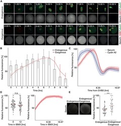
- FIGURE 1 The expression and proteolysis of endogenous and exogenous securin during meiosis I. (A) Upper panel shows representative images of fixed oocytes from indicated time intervals after IBMX removal. DNA (red) was visualized by DAPI, tubulin (green) and securin (gray) were detected by anti-tubulin and anti-securin antibodies. Scale bar represents 10 mum. Lower panel shows representative time frames from live cell imaging of oocytes microinjected with cRNAs encoding histone H2B (red), tubulin (green), and securin (gray) fused to fluorescent proteins. Scale bar represents 20 mum. (B) Expression profiles of endogenous securin (columns, 0 h n = 26, 1 h n = 25, 2 h n = 26, 3 h n = 26, 4 h n = 26, 5 h n = 26, 6 h n = 26, 7 h n = 26, 8 h n = 25, 9 h n = 21, and 10 h n = 24) and exogenous securin (red line, all time intervals n = 37). Time from IBMX removal. The data for endogenous securin were obtained from two independent experiments and for exogenous securin from three independent experiments. (C) Fluorescence signal profiles of securin (red, n = 28) and cyclin A2 (blue, n = 28) during meiosis I. Time is from GVBD. The data were obtained from three independent experiments. (D) Left panel shows scatterplot of the endogenous securin signal in oocytes at 0 h (mean: 63.01%, n = 78) and at 12 h (mean: 59.84%, n = 78) of IBMX treatment. The difference between groups was not statistically significant (alpha > 0.05, n.s. P = 0.2554). The data were obtained from three independent expe

- Submitted by
- Invitrogen Antibodies (provider)
- Main image

- Experimental details
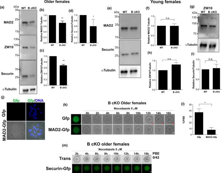
- FIGURE 4 Aurkb cKO oocytes from older females have reduced expression of MAD2, ZW10 and Securin. (a) Representative Western blot images of prophase I-arrested oocytes from older wild-type (WT) or conditional Aurkb knockout (B cKO) females detecting MAD2, ZW10 and Securin. (b) Quantification of MAD2 (** p < 0.0030), (c) Quantification of ZW10 (** p < 0.0016). (d) Quantification of Securin (* p < 0.0468). (e, g) Representative Western blots of prophase I-arrested oocytes from young females detecting MAD2, Securin (d) and ZW10 (g). (f) Quantification of MAD2 ( p = 0.9774). Graph shows individual values plus the mean +-SEM from 3 independent experiments. (h) Quantification of ZW10 ( p = 0.1166). (i) Quantification of Securin (* p < 0.7505), MAD2, ZW10 and Securin signals were normalized to alpha-Tubulin. Each lane contained 100 prophase I-arrested oocytes. n.s. : not significant. Graph shows mean +-SEM from 2-3 independent experiments. (j) Representative confocal images of pro-metaphase I oocytes expressing Gfp or MAD2-gfp (green) DNA (blue). Scale bar: 10 mum. (k) Representative images of timing of PBE of Aurkb cKO oocytes from older females, matured in nocodazole expressing either Gfp or MAD2-gfp. Red star indicates a PB. Scale bar: 20 mum. (l) Quantification of % of oocytes from (k) that undergo PBE in nocodazole. (* p = 0.0.0160; number of oocytes examined Gfp: 73, MAD2-gfp: 59; 6 mice). Graph shows the mean +-SEM from 3 experiments. (m) Representative images of timing of PBE