Explore
 Validate
 Learn
Learn Western blot
 Immunocytochemistry
Antibody data
- Antibody Data
- Antigen structure
- References [1]
- Comments [0]
- Validations
- Immunocytochemistry [3]
- Immunoprecipitation [1]
- Immunohistochemistry [1]
- Other assay [2]
Submit
Validation data
Reference
Comment
Report error
- Product number
- PA5-29399 - Provider product page

- Provider
- Invitrogen Antibodies
- Product name
- Securin Polyclonal Antibody
- Antibody type
- Polyclonal
- Antigen
- Recombinant full-length protein
- Description
- Recommended positive controls: IMR32. Predicted reactivity: Rhesus Monkey (98%), Chimpanzee (99%), Bovine (85%). Store product as a concentrated solution. Centrifuge briefly prior to opening the vial.
- Reactivity
- Human
- Host
- Rabbit
- Isotype
- IgG
- Vial size
- 100 μL
- Concentration
- 1 mg/mL
- Storage
- Store at 4°C short term. For long term storage, store at -20°C, avoiding freeze/thaw cycles.
Submitted references Signatures of Multi-Omics Reveal Distinct Tumor Immune Microenvironment Contributing to Immunotherapy in Lung Adenocarcinoma.
Huang Z, Li B, Guo Y, Wu L, Kou F, Yang L
Frontiers in immunology 2021;12:723172
Frontiers in immunology 2021;12:723172
No comments: Submit comment
Supportive validation
- Submitted by
- Invitrogen Antibodies (provider)
- Main image
- Experimental details
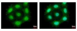
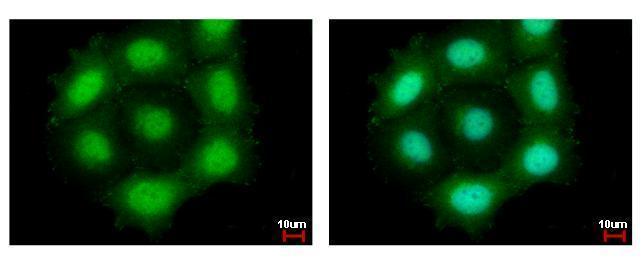
- Securin Polyclonal Antibody detects PTTG1 protein at cytoplasm and nucleus by immunofluorescent analysis. Sample: MCF-7 cells were fixed in 4% paraformaldehyde at RT for 15 min. Green: PTTG1 protein stained by Securin Polyclonal Antibody (Product # PA5-29399) diluted at 1:500. Blue: Hoechst 33342 staining.

- Submitted by
- Invitrogen Antibodies (provider)
- Main image

- Experimental details
- Securin Polyclonal Antibody detects PTTG1 protein at cytoplasm and nucleus by immunofluorescent analysis. Sample: MCF-7 cells were fixed in 4% paraformaldehyde at RT for 15 min. Green: PTTG1 protein stained by Securin Polyclonal Antibody (Product # PA5-29399) diluted at 1:500. Blue: Hoechst 33342 staining.

Supportive validation
- Submitted by
- Invitrogen Antibodies (provider)
- Main image
- Experimental details
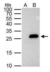
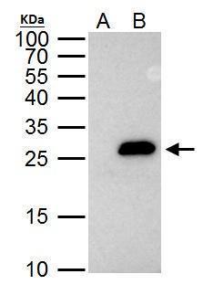
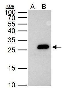
- Securin Polyclonal Antibody immunoprecipitates Securin protein in IP experiments. IP Sample: HeLa whole cell lysate/extract A. Control with 2 µg of preimmune rabbit IgG B. Immunoprecipitation of Securin protein by 2 µg of Securin Polyclonal Antibody (Product # PA5-29399) 12% SDS-PAGE The immunoprecipitated Securin protein was detected by Securin Polyclonal Antibody (Product # PA5-29399) diluted at 1:1,000.
Supportive validation
- Submitted by
- Invitrogen Antibodies (provider)
- Main image
- Experimental details
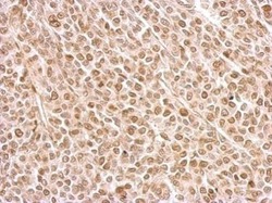
- Securin Polyclonal Antibody detects PTTG1 protein at on BT483 xenograft by immunohistochemical analysis. Sample: Paraffin-embedded BT483 xenograft. Securin Polyclonal Antibody (Product # PA5-29399) dilution: 1:500. Antigen Retrieval: EDTA based buffer, pH 8.0, 15 min.
Supportive validation
- Submitted by
- Invitrogen Antibodies (provider)
- Main image
- Experimental details
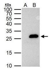
- Securin Polyclonal Antibody immunoprecipitates Securin protein in IP experiments. IP Sample: HeLa whole cell lysate/extract A. Control with 2 µg of preimmune rabbit IgG B. Immunoprecipitation of Securin protein by 2 µg of Securin Polyclonal Antibody (Product # PA5-29399) 12% SDS-PAGE The immunoprecipitated Securin protein was detected by Securin Polyclonal Antibody (Product # PA5-29399) diluted at 1:1,000.
- Submitted by
- Invitrogen Antibodies (provider)
- Main image
- Experimental details
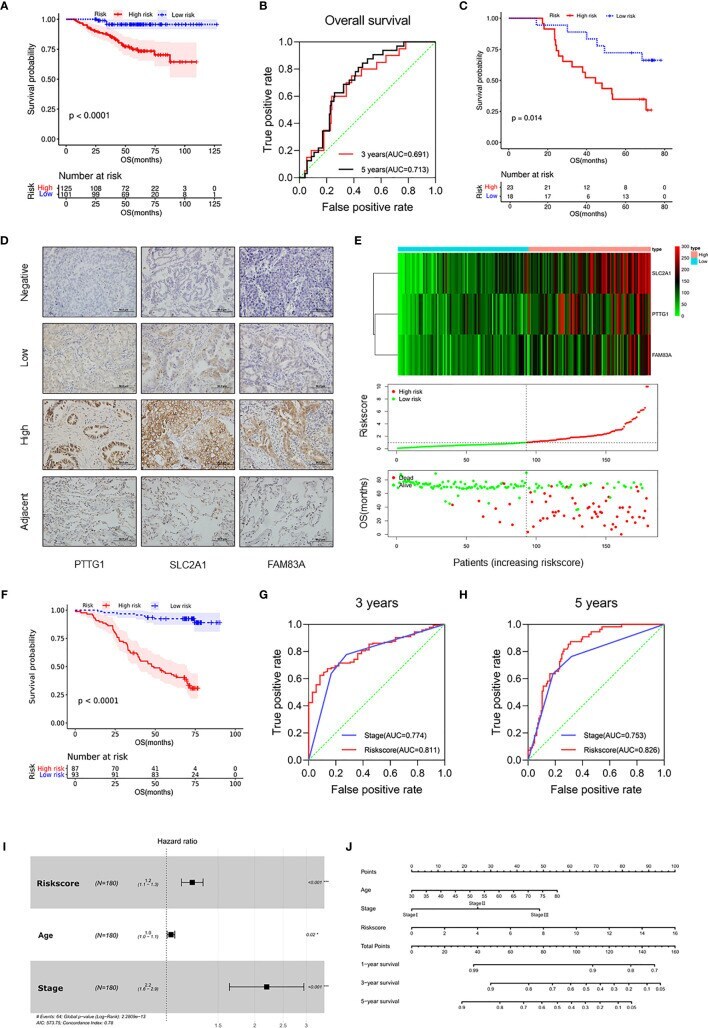
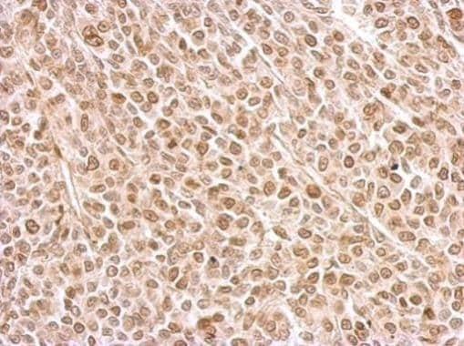
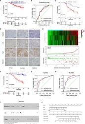
- Figure 4 Validation of the prognostic risk model in the LUAD cohort. (A) Kaplan-Meier curves for OS by risk subgroups in the GSE31210. (B) Time-dependent ROC curves measuring the prognostic value of the risk score at 3 and 5 years in the GSE31210. (C) Kaplan-Meier curves for OS by risk subgroups in the mRNA level from the Tianjin cohort. (D) Representative IHC images for staining of signatures in 180 samples of patients with LUAD (Olympus, x400 magnification, bar = 50 mum). (E) Heat map of PTTG1 , SLC2A1 , and FAM83A in the Tianjin cohort (upper figure). Distribution of risk score (middle figure), and OS (lower figure). (F) Kaplan-Meier curves for OS by risk subgroups in the Tianjin cohort. (G, H) Time-dependent ROC curves measuring the prognostic value of the risk score and stage at (G) 3 years and (H) 5 years in the Tianjin cohort. (I) Forest plot showing the independent prognostic factors for OS in the Tianjin cohort (multivariate Cox regression analysis). (J) Nomogram for predicting 1-, 3-, and 5-year overall survival in the Tianjin cohort. *p < 0.05 and ***p < 0.001.