Explore
 Validate
 Learn
Learn Western blot
Antibody data
- Antibody Data
- Antigen structure
- References [3]
- Comments [0]
- Validations
- Western blot [1]
- Immunohistochemistry [1]
Submit
Validation data
Reference
Comment
Report error
- Product number
- sc-11429 - Provider product page

- Provider
- Santa Cruz Biotechnology
- Proper citation
- Santa Cruz Biotechnology Cat#sc-11429, RRID:AB_2215661
- Product name
- Anti-XRCC1
- Antibody type
- Polyclonal
- Antigen
- Recombinant full-length protein
- Reactivity
- Human
- Host
- Rabbit
Submitted references Senescence evasion by MCF-7 human breast tumor-initiating cells.
Dual functions of ASCIZ in the DNA base damage response and pulmonary organogenesis.
Hsp70 translocates to the nuclei and nucleoli, binds to XRCC1 and PARP-1, and protects HeLa cells from single-strand DNA breaks.
Karimi-Busheri F, Rasouli-Nia A, Mackey JR, Weinfeld M
Breast cancer research : BCR 2010;12(3):R31
Breast cancer research : BCR 2010;12(3):R31
Dual functions of ASCIZ in the DNA base damage response and pulmonary organogenesis.
Jurado S, Smyth I, van Denderen B, Tenis N, Hammet A, Hewitt K, Ng JL, McNees CJ, Kozlov SV, Oka H, Kobayashi M, Conlan LA, Cole TJ, Yamamoto K, Taniguchi Y, Takeda S, Lavin MF, Heierhorst J
PLoS genetics 2010 Oct 21;6(10):e1001170
PLoS genetics 2010 Oct 21;6(10):e1001170
Hsp70 translocates to the nuclei and nucleoli, binds to XRCC1 and PARP-1, and protects HeLa cells from single-strand DNA breaks.
Kotoglou P, Kalaitzakis A, Vezyraki P, Tzavaras T, Michalis LK, Dantzer F, Jung JU, Angelidis C
Cell stress & chaperones 2009 Jul;14(4):391-406
Cell stress & chaperones 2009 Jul;14(4):391-406
No comments: Submit comment
Supportive validation
- Submitted by
- per
- Main image

- Experimental details
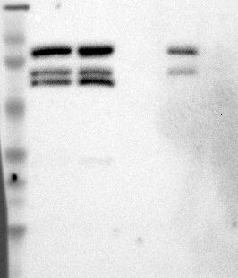
- Western blot analysis of antibody specificity using a routine panel composed of IgG/HSA-depleted human plasma and protein lysates from selected human tissues and cell lines.
- Validation comment
- Band of predicted size in kDa (+/-20%) with additional bands present.
- Primary Ab dilution
- 1:500
- Secondary Ab dilution
- 1:3000
- Lane 1
- Marker [kDa]: 229, 112, 83.5, 47.9, 32.3, 26.5, 17.2
- Lane 2
- RT-4
- Lane 3
- U-251MG sp
- Lane 4
- Human Plasma
- Lane 5
- Liver
- Lane 6
- Tonsil
- Theoretical target weight
- [kDa] 69
Supportive validation
- Submitted by
- per
- Main image
- Experimental details
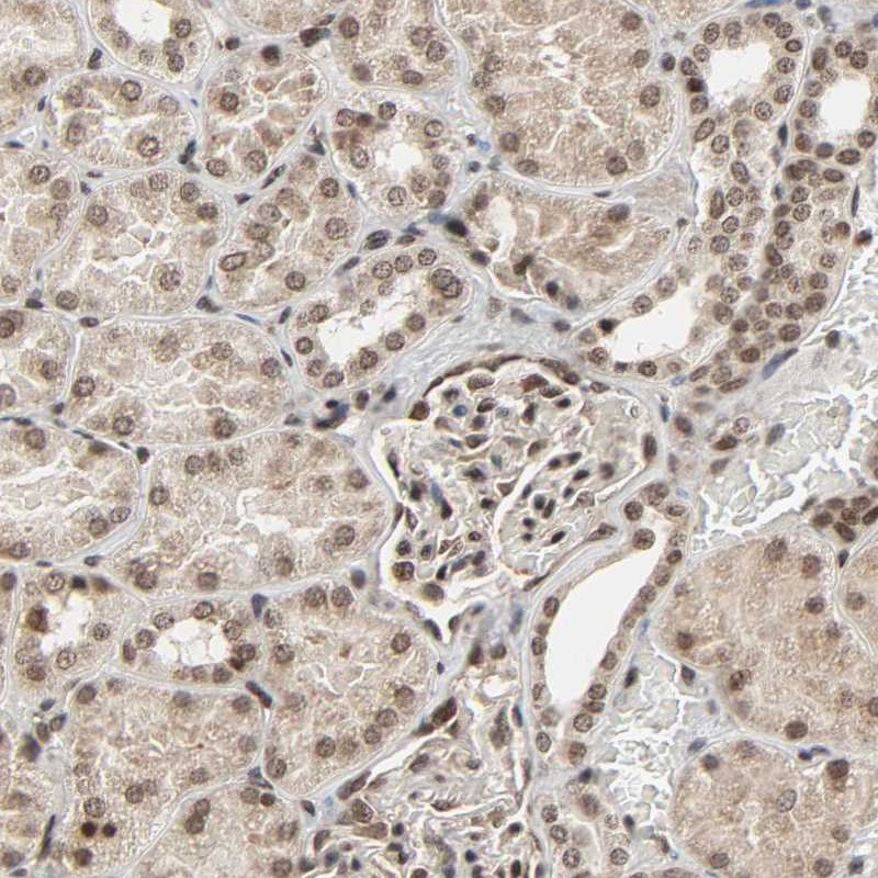
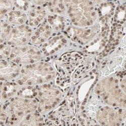
- Immunohistochemical staining of human kidney shows moderate nuclear positivity in cells in tubules.
- Validation comment
- Staining pattern consistent with experimental and/or bioinformatic data.