Explore
 Validate
 Learn
Learn Western blot
 Immunoprecipitation
Antibody data
- Antibody Data
- Antigen structure
- References [26]
- Comments [0]
- Validations
- Western blot [1]
- Immunohistochemistry [2]
- Other assay [5]
Submit
Validation data
Reference
Comment
Report error
- Product number
- MA5-13412 - Provider product page

- Provider
- Invitrogen Antibodies
- Product name
- XRCC1 Monoclonal Antibody (33-2-5)
- Antibody type
- Monoclonal
- Antigen
- Recombinant full-length protein
- Description
- MA5-13412 targets XRCC1 in IHC (P), IP, and WB applications and shows reactivity with Human and Rat samples. The MA5-13412 immunogen is recombinant human XRCC1 protein.
- Reactivity
- Human, Rat
- Host
- Mouse
- Isotype
- IgG
- Antibody clone number
- 33-2-5
- Vial size
- 500 µL
- Concentration
- 0.2 mg/mL
- Storage
- 4° C
Submitted references The Novel Role of hnRNP UL1 in Human Cell Nucleoli.
Linker region is required for efficient nuclear localization of polynucleotide kinase phosphatase.
Ligand-induced gene activation is associated with oxidative genome damage whose repair is required for transcription.
Defective base excision repair in the response to DNA damaging agents in triple negative breast cancer.
Base excision repair defects invoke hypersensitivity to PARP inhibition.
Interaction between DNA Polymerase β and BRCA1.
Preventing oxidation of cellular XRCC1 affects PARP-mediated DNA damage responses.
Targeting XRCC1 deficiency in breast cancer for personalized therapy.
Clinicopathological and functional significance of XRCC1 expression in ovarian cancer.
The Concept of Divergent Targeting through the Activation and Inhibition of Receptors as a Novel Chemotherapeutic Strategy: Signaling Responses to Strong DNA-Reactive Combinatorial Mimicries.
E2F1 is involved in DNA single-strand break repair through cell-cycle-dependent upregulation of XRCC1 expression.
The prognostic significance of ERCC1, BRCA1, XRCC1, and betaIII-tubulin expression in patients with non-small cell lung cancer treated by platinum- and taxane-based neoadjuvant chemotherapy and surgical resection.
Subcellular distribution of a fluorescence-labeled combi-molecule designed to block epidermal growth factor receptor tyrosine kinase and damage DNA with a green fluorescent species.
XRCC1 protects against the lethality of induced oxidative DNA damage in nondividing neural cells.
Chk2-dependent phosphorylation of XRCC1 in the DNA damage response promotes base excision repair.
Tamoxifen accelerates proteasomal degradation of O6-methylguanine DNA methyltransferase in human cancer cells.
Human Xip1 (C2orf13) is a novel regulator of cellular responses to DNA strand breaks.
An intronic polymorphism associated with increased XRCC1 expression, reduced apoptosis and familial breast cancer.
Replication-dependent and -independent responses of RAD18 to DNA damage in human cells.
XRCC1 and DNA polymerase beta interaction contributes to cellular alkylating-agent resistance and single-strand break repair.
The Arg280His polymorphism in X-ray repair cross-complementing gene 1 impairs DNA repair ability.
In situ analysis of repair processes for oxidative DNA damage in mammalian cells.
Involvement of poly(ADP-ribose) polymerase-1 and XRCC1/DNA ligase III in an alternative route for DNA double-strand breaks rejoining.
X-ray repair cross-complementing gene I protein plays an important role in camptothecin resistance.
Oxidative DNA damage and repair in experimental atherosclerosis are reversed by dietary lipid lowering.
Oxidative DNA damage and repair in experimental atherosclerosis are reversed by dietary lipid lowering.
Cichocka M, Karlik A, Plewka P, Gawade K, Stępień A, Świergiel P, Gadgil A, Raczyńska KD
International journal of biological sciences 2022;18(13):4809-4823
International journal of biological sciences 2022;18(13):4809-4823
Linker region is required for efficient nuclear localization of polynucleotide kinase phosphatase.
Tsukada K, Matsumoto Y, Shimada M
PloS one 2020;15(9):e0239404
PloS one 2020;15(9):e0239404
Ligand-induced gene activation is associated with oxidative genome damage whose repair is required for transcription.
Sengupta S, Wang H, Yang C, Szczesny B, Hegde ML, Mitra S
Proceedings of the National Academy of Sciences of the United States of America 2020 Sep 8;117(36):22183-22192
Proceedings of the National Academy of Sciences of the United States of America 2020 Sep 8;117(36):22183-22192
Defective base excision repair in the response to DNA damaging agents in triple negative breast cancer.
Lee KJ, Piett CG, Andrews JF, Mann E, Nagel ZD, Gassman NR
PloS one 2019;14(10):e0223725
PloS one 2019;14(10):e0223725
Base excision repair defects invoke hypersensitivity to PARP inhibition.
Horton JK, Stefanick DF, Prasad R, Gassman NR, Kedar PS, Wilson SH
Molecular cancer research : MCR 2014 Aug;12(8):1128-39
Molecular cancer research : MCR 2014 Aug;12(8):1128-39
Interaction between DNA Polymerase β and BRCA1.
Masaoka A, Gassman NR, Horton JK, Kedar PS, Witt KL, Hobbs CA, Kissling GE, Tano K, Asagoshi K, Wilson SH
PloS one 2013;8(6):e66801
PloS one 2013;8(6):e66801
Preventing oxidation of cellular XRCC1 affects PARP-mediated DNA damage responses.
Horton JK, Stefanick DF, Gassman NR, Williams JG, Gabel SA, Cuneo MJ, Prasad R, Kedar PS, Derose EF, Hou EW, London RE, Wilson SH
DNA repair 2013 Sep;12(9):774-85
DNA repair 2013 Sep;12(9):774-85
Targeting XRCC1 deficiency in breast cancer for personalized therapy.
Sultana R, Abdel-Fatah T, Abbotts R, Hawkes C, Albarakati N, Seedhouse C, Ball G, Chan S, Rakha EA, Ellis IO, Madhusudan S
Cancer research 2013 Mar 1;73(5):1621-34
Cancer research 2013 Mar 1;73(5):1621-34
Clinicopathological and functional significance of XRCC1 expression in ovarian cancer.
Abdel-Fatah T, Sultana R, Abbotts R, Hawkes C, Seedhouse C, Chan S, Madhusudan S
International journal of cancer 2013 Jun 15;132(12):2778-86
International journal of cancer 2013 Jun 15;132(12):2778-86
The Concept of Divergent Targeting through the Activation and Inhibition of Receptors as a Novel Chemotherapeutic Strategy: Signaling Responses to Strong DNA-Reactive Combinatorial Mimicries.
Watt HL, Rachid Z, Jean-Claude BJ
Journal of signal transduction 2012;2012:282050
Journal of signal transduction 2012;2012:282050
E2F1 is involved in DNA single-strand break repair through cell-cycle-dependent upregulation of XRCC1 expression.
Jin R, Sun Y, Qi X, Zhang H, Zhang Y, Li N, Ding W, Chen D
DNA repair 2011 Sep 5;10(9):926-33
DNA repair 2011 Sep 5;10(9):926-33
The prognostic significance of ERCC1, BRCA1, XRCC1, and betaIII-tubulin expression in patients with non-small cell lung cancer treated by platinum- and taxane-based neoadjuvant chemotherapy and surgical resection.
Kang CH, Jang BG, Kim DW, Chung DH, Kim YT, Jheon S, Sung SW, Kim JH
Lung cancer (Amsterdam, Netherlands) 2010 Jun;68(3):478-83
Lung cancer (Amsterdam, Netherlands) 2010 Jun;68(3):478-83
Subcellular distribution of a fluorescence-labeled combi-molecule designed to block epidermal growth factor receptor tyrosine kinase and damage DNA with a green fluorescent species.
Todorova MI, Larroque AL, Dauphin-Pierre S, Fang YQ, Jean-Claude BJ
Molecular cancer therapeutics 2010 Apr;9(4):869-82
Molecular cancer therapeutics 2010 Apr;9(4):869-82
XRCC1 protects against the lethality of induced oxidative DNA damage in nondividing neural cells.
Kulkarni A, McNeill DR, Gleichmann M, Mattson MP, Wilson DM 3rd
Nucleic acids research 2008 Sep;36(15):5111-21
Nucleic acids research 2008 Sep;36(15):5111-21
Chk2-dependent phosphorylation of XRCC1 in the DNA damage response promotes base excision repair.
Chou WC, Wang HC, Wong FH, Ding SL, Wu PE, Shieh SY, Shen CY
The EMBO journal 2008 Dec 3;27(23):3140-50
The EMBO journal 2008 Dec 3;27(23):3140-50
Tamoxifen accelerates proteasomal degradation of O6-methylguanine DNA methyltransferase in human cancer cells.
Kuo CC, Liu JF, Shiah HS, Ma LC, Chang JY
International journal of cancer 2007 Nov 15;121(10):2293-300
International journal of cancer 2007 Nov 15;121(10):2293-300
Human Xip1 (C2orf13) is a novel regulator of cellular responses to DNA strand breaks.
Bekker-Jensen S, Fugger K, Danielsen JR, Gromova I, Sehested M, Celis J, Bartek J, Lukas J, Mailand N
The Journal of biological chemistry 2007 Jul 6;282(27):19638-43
The Journal of biological chemistry 2007 Jul 6;282(27):19638-43
An intronic polymorphism associated with increased XRCC1 expression, reduced apoptosis and familial breast cancer.
Bu D, Tomlinson G, Lewis CM, Zhang C, Kildebeck E, Euhus DM
Breast cancer research and treatment 2006 Oct;99(3):257-65
Breast cancer research and treatment 2006 Oct;99(3):257-65
Replication-dependent and -independent responses of RAD18 to DNA damage in human cells.
Nakajima S, Lan L, Kanno S, Usami N, Kobayashi K, Mori M, Shiomi T, Yasui A
The Journal of biological chemistry 2006 Nov 10;281(45):34687-95
The Journal of biological chemistry 2006 Nov 10;281(45):34687-95
XRCC1 and DNA polymerase beta interaction contributes to cellular alkylating-agent resistance and single-strand break repair.
Wong HK, Wilson DM 3rd
Journal of cellular biochemistry 2005 Jul 1;95(4):794-804
Journal of cellular biochemistry 2005 Jul 1;95(4):794-804
The Arg280His polymorphism in X-ray repair cross-complementing gene 1 impairs DNA repair ability.
Takanami T, Nakamura J, Kubota Y, Horiuchi S
Mutation research 2005 Apr 4;582(1-2):135-45
Mutation research 2005 Apr 4;582(1-2):135-45
In situ analysis of repair processes for oxidative DNA damage in mammalian cells.
Lan L, Nakajima S, Oohata Y, Takao M, Okano S, Masutani M, Wilson SH, Yasui A
Proceedings of the National Academy of Sciences of the United States of America 2004 Sep 21;101(38):13738-43
Proceedings of the National Academy of Sciences of the United States of America 2004 Sep 21;101(38):13738-43
Involvement of poly(ADP-ribose) polymerase-1 and XRCC1/DNA ligase III in an alternative route for DNA double-strand breaks rejoining.
Audebert M, Salles B, Calsou P
The Journal of biological chemistry 2004 Dec 31;279(53):55117-26
The Journal of biological chemistry 2004 Dec 31;279(53):55117-26
X-ray repair cross-complementing gene I protein plays an important role in camptothecin resistance.
Park SY, Lam W, Cheng YC
Cancer research 2002 Jan 15;62(2):459-65
Cancer research 2002 Jan 15;62(2):459-65
Oxidative DNA damage and repair in experimental atherosclerosis are reversed by dietary lipid lowering.
Martinet W, Knaapen MW, De Meyer GR, Herman AG, Kockx MM
Circulation research 2001 Apr 13;88(7):733-9
Circulation research 2001 Apr 13;88(7):733-9
Oxidative DNA damage and repair in experimental atherosclerosis are reversed by dietary lipid lowering.
Martinet W, Knaapen MW, De Meyer GR, Herman AG, Kockx MM
Circulation research 2001 Apr 13;88(7):733-9
Circulation research 2001 Apr 13;88(7):733-9
No comments: Submit comment
Supportive validation
- Submitted by
- Invitrogen Antibodies (provider)
- Main image
- Experimental details
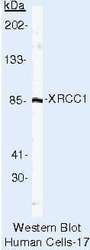
- Western blot of XRCC1 using XRCC1 Monoclonal Antibody (Product # MA5-13412) on LS174T Cells.
Supportive validation
- Submitted by
- Invitrogen Antibodies (provider)
- Main image
- Experimental details
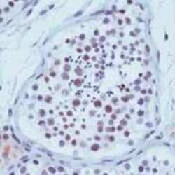
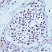
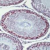
- Formalin-fixed, paraffin-embedded human testis stained with XRCC1 antibody using peroxidase-conjugate and AEC chromogen. Note nuclear staining of spermatocytes.
- Submitted by
- Invitrogen Antibodies (provider)
- Main image
- Experimental details
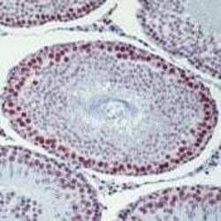
- Formalin-fixed, paraffin-embedded rat testis stained with XRCC1 antibody using peroxidase-conjugate and AEC chromogen. Note nuclear staining of spermatocytes.
Supportive validation
- Submitted by
- Invitrogen Antibodies (provider)
- Main image
- Experimental details
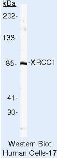
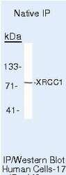
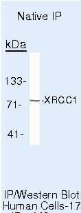
- Immunoprecipitation of XRCC1 using XRCC1 Monoclonal Antibody (Product # MA5-13412) on Native Human LS174T Cells.
- Submitted by
- Invitrogen Antibodies (provider)
- Main image
- Experimental details
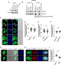
- Fig 5 FHA domain of PNKP is involved in distribution to subnuclear compartments. (A) Confirmation of protein expression of GFP-PNKP constructs in U2OS cells by western blot analysis. (B) Interaction between FL PNKP and PNKP mutants (R35A, K138A and R35A/K138A) examined by immunoprecipitation. GFP-Trap magnetic agarose beads were used for immunoprecipitation from GFP-tagged PNKP expressing HEK293 cells. XRCC1 and XRCC4 were detected by western blotting and GFP-tagged PNKP was detected by Ponceau staining. (C) Representative live-cell images of GFP-tagged PNKP mutants, expressing in U2OS cells, with DIC images. Scale bar indicates as 10mum. (D, E, F) Dot plot of the N/C ratio of GFI. At least 150 cells were analyzed and plotted. (G) GFP-tagged FL PNKP and R35A PNKP localization in nucleus. Subnuclear regions with increased or decreased GFI are surrounded by white or black dashed circles, respectively. (H) Representative live-cell images of GFP-tagged FL PNKP, R35A PNKP and mCherry-tagged Fibrillarin localization in nucleolus. (I) Dot plot of the nucleous/nucleus ratio of GFI. At least 150 cells were analyzed and plotted. ns: not significant (p > 0.05); **: 0.005 < p
- Submitted by
- Invitrogen Antibodies (provider)
- Main image
- Experimental details
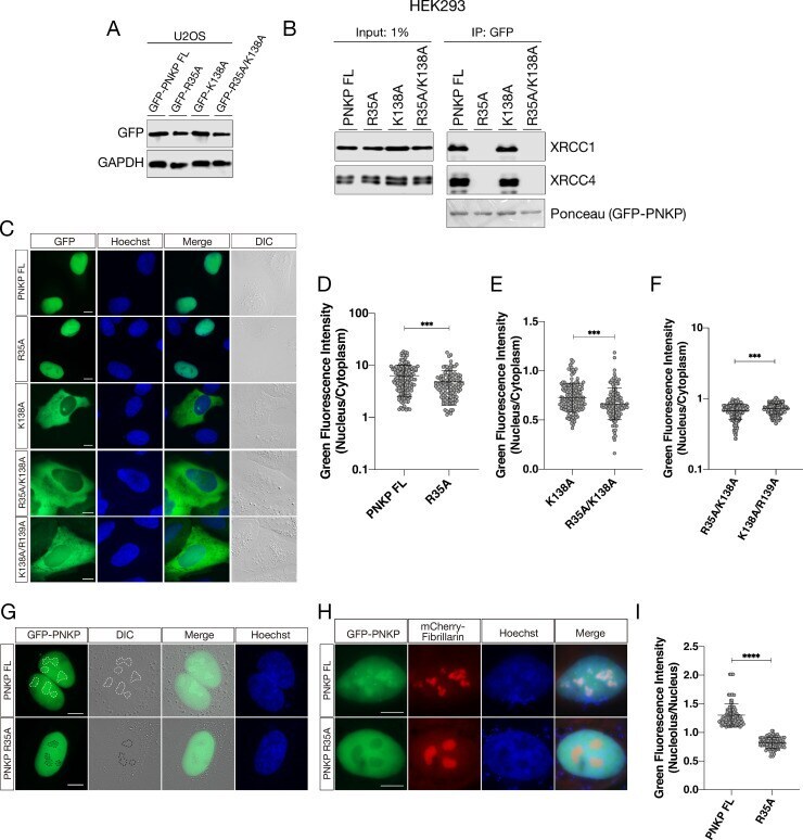
- Involvement of hnRNP UL1 in the DDR in human nucleoli. (A) The DNA damage sensitivity of HEK UL1 KO cells compared to HEK WT cells was tested after 2.5 h of treatment with the genotoxic reagents ETO and CPT. After this time, the cells were harvested at the following time points: 18 h, 24 h, 30 h, 48 h, 72 h and 80 h. Cell viability was assessed by Trypan blue staining, and the percentage of survival was calculated. The results were normalized to those for the control cells treated with DMSO. (B) DNA damage was assessed by Comet Assay in HEK UL1 KO cells compared to HEK WT cells after DNA damage induced with ETO and CPT. Cells treated with DMSO were used as negative controls. For the assay, 50 cells per sample were analyzed. The error bars represent the SDs of three biological replicates. The p values were calculated using Student's t test, and the statistical significance is represented as follows: *P

- Submitted by
- Invitrogen Antibodies (provider)
- Main image
- Experimental details
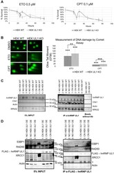
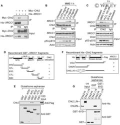
- Interaction between Chk2 and XRCC1. ( A ) In vivo association of Chk2 with XRCC1. 293T cells were co-transfected with Myc-tagged Chk2 and/or His-tagged XRCC1 , then whole-cell extracts were subjected to immunoprecipitation (IP) with anti-His antibody and the immunoprecipitated proteins analysed by immunoblotting (IB) with anti-Myc or anti-His antibodies (upper panel). The lower panel shows IB of the whole-cell extract. ( B ) Association of Chk2 and XRCC1 is damage-inducible. 293T cells were either untreated or treated for 1 h with increasing concentrations of methyl methanesulphonate (MMS), then whole-cell lysates were subjected to IP with anti-Chk2 antibody and the immunoprecipitated proteins were analysed on IB using anti-XRCC1 or anti-Chk2 antibodies (upper panel). The lower panel shows IB of the whole-cell extract for Chk2, XRCC1, and phosphorylated p53 (p53-pS15), an indicator of DNA damage. ( C ) In vivo association of endogenous Chk2 with endogenous XRCC1 is induced by alkylation (MMS), oxidative damage (H 2 O 2 ), or ionizing radiation (IR) (upper panel). Whole-cell lysates were analysed in the lower panel as in (B). ( D ) Constructs of GST-fused XRCC1 segments used in the in vitro binding assay. ( E ) Upper panel, purified Flag-tagged Chk2 produced in bacteria was incubated with purified GST-XRCC1 fragments, and the proteins pulled down by glutathione beads were analysed by IB with anti-Flag antibody. Lower panel, pull down of GST-XRCC1 fusion proteins was confirmed
- Submitted by
- Invitrogen Antibodies (provider)
- Main image
- Experimental details
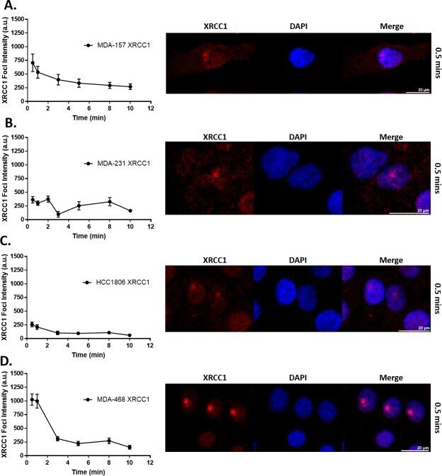
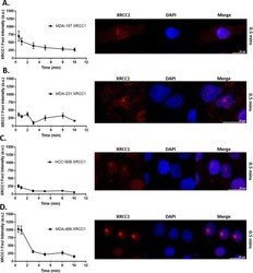
- Fig 6 XRCC1 recruitment following 355 nm laser microirradiation in TNBC cell lines. Microirradiation with a 355 nm laser occurred and cells were fixed at the indicated time points and processed for immunofluorescence of XRCC1. Quantification over time is shown on the left with a representative image from 0.5 min shown on the right for A) MDA-157, B) MDA-231, C) HCC1806, and D) MDA-468. A minimum of 18 cells were irradiated over three separate experiments and quantified as described in the materials and methods section and reported as XRCC1 Foci Intensity in arbitrary units (a.u.). Scale bar = 20 mum.