Explore
 Validate
 Learn
Learn Western blot
Antibody data
- Antibody Data
- Antigen structure
- References [0]
- Comments [0]
- Validations
- Western blot [2]
- Immunocytochemistry [1]
- Immunohistochemistry [1]
Submit
Validation data
Reference
Comment
Report error
- Product number
- ASC-031-25UL - Provider product page

- Provider
- Invitrogen Antibodies
- Product name
- pan ASIC (extracellular) Polyclonal Antibody
- Antibody type
- Polyclonal
- Antigen
- Other
- Reactivity
- Human, Mouse, Rat
- Host
- Rabbit
- Isotype
- IgG
- Vial size
- 25 µL
- Concentration
- 0.8 mg/mL
- Storage
- -20° C, Avoid Freeze/Thaw Cycles
No comments: Submit comment
Supportive validation
- Submitted by
- Invitrogen Antibodies (provider)
- Main image
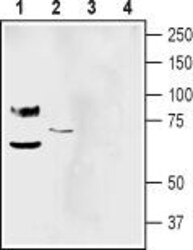
- Experimental details
- Western blot analysis of mouse brain membranes (lanes 1 and 3) and human SH-SY5Y neuroblastoma cell line lysate (lanes 2 and 4): - 1-2. Anti-pan ASIC (extracellular) Antibody (#ASC-031), (1:500).3-4. Anti-pan ASIC (extracellular) Antibody , preincubated with Pan ASIC (extracellular) Blocking Peptide (#BLP-SC031).
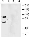
- Submitted by
- Invitrogen Antibodies (provider)
- Main image

- Experimental details
- Western blot analysis of rat dorsal root ganglion lysate (lanes 1 and 3) and rat brain membranes (lanes 2 and 4): - 1-2. Anti-pan ASIC (extracellular) Antibody (#ASC-031), (1:500).3-4. Anti-pan ASIC (extracellular) Antibody , preincubated with Pan ASIC (extracellular) Blocking Peptide (#BLP-SC031).
Supportive validation
- Submitted by
- Invitrogen Antibodies (provider)
- Main image
- Experimental details
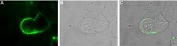
- Expression of ASIC in rat PC12 cells - Cell surface detection of ASIC in live intact rat PC12 pheochromocytoma cells. A. Extracellular staining of cells with Anti-pan ASIC (extracellular) Antibody (#ASC-031), (1:25), followed by goat Anti-rabbit-AlexaFluor-488 secondary Antibody (green). B. Live view of the cells. C. Merge of A and B.

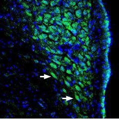
Supportive validation
- Submitted by
- Invitrogen Antibodies (provider)
- Main image
- Experimental details
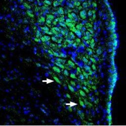
- Expression of ASIC channels in rat locus coeruleus - Immunohistochemical staining of perfusion-fixed rat brain frozen sections using Anti-pan ASIC (extracellular) Antibody (#ASC-031), (1:400), followed by goat- Anti-rabbit-AlexaFluor-488. ASIC channel staining (green) appears in neuronal soma (arrows). Nuclei are stained with DAPI (blue).