Explore
 Validate
 Learn
Learn Western blot
 Flow cytometry
Antibody data
- Antibody Data
- Antigen structure
- References [2]
- Comments [0]
- Validations
- Western blot [1]
Submit
Validation data
Reference
Comment
Report error
- Product number
- PA1938 - Provider product page

- Provider
- Boster Biological Technology
- Product name
- Anti-Acid-sensing ion channel 3 ASIC3 Antibody
- Antibody type
- Polyclonal
- Description
- Polyclonal antibody for ASIC3 detection. Host: Rabbit.Size: 100μg/vial. Tested applications: WB. Reactive species: Human. ASIC3 information: Molecular Weight: 58905 MW; Subcellular Localization: Cell membrane ; Multi-pass membrane protein . Cytoplasm . Cell surface expression may be stabilized by interaction with LIN7B and cytoplasmic retention by interaction with DLG4. In part cytoplasmic in cochlea cells (By similarity); Tissue Specificity: Expressed by sensory neurons. Strongly expressed in brain, spinal chord, lung, lymph nodes, kidney, pituitary, heart and testis.
- Reactivity
- Human, Mouse, Rat
- Host
- Rabbit
- Vial size
- 100μg/vial
- Concentration
- Add 0.2ml of distilled water will yield a concentration of 500ug/ml.
- Storage
- At -20°C for one year. After reconstitution, at 4°C for one month. It can also be aliquoted and stored frozen at -20°C for a longer time. Avoid repeated freezing and thawing.
- Handling
- Add 0.2ml of distilled water will yield a concentration of 500ug/ml.
Submitted references Effect of knockout of the ASIC3 on cardiovascular reflexes arising from hindlimb muscle in decerebrated rats.
Naked mole-rat acid-sensing ion channel 3 forms nonfunctional homomers, but functional heteromers.
Kim JS, Harms JE, Ruiz-Velasco V, Kaufman MP
American journal of physiology. Regulatory, integrative and comparative physiology 2019 Nov 1;317(5):R641-R648
American journal of physiology. Regulatory, integrative and comparative physiology 2019 Nov 1;317(5):R641-R648
Naked mole-rat acid-sensing ion channel 3 forms nonfunctional homomers, but functional heteromers.
Schuhmacher LN, Callejo G, Srivats S, Smith ESJ
The Journal of biological chemistry 2018 Feb 2;293(5):1756-1766
The Journal of biological chemistry 2018 Feb 2;293(5):1756-1766
No comments: Submit comment
Supportive validation
- Submitted by
- Boster Biological Technology (provider)
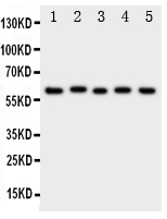
- Main image
- Experimental details
- Western blot analysis of ASIC3 using anti- ASIC3 antibody (PA1938). Electrophoresis was performed on a 5-20% SDS-PAGE gel at 70V (Stacking gel) / 90V (Resolving gel) for 2-3 hours. The sample well of each lane was loaded with 50ug of sample under reducing conditions. Lane 1: Rat Brain Tissue Lysate, Lane 2: Rat Testis Tissue Lysate, Lane 3: U87 Cell Lysate, Lane 4: NEURO Cell Lysate, Lane 5: SMMC Cell Lysate. After Electrophoresis, proteins were transferred to a Nitrocellulose membrane at 150mA for 50-90 minutes. Blocked the membrane with 5% Non-fat Milk/ TBS for 1.5 hour at RT. The membrane was incubated with rabbit anti- ASIC3 antigen affinity purified polyclonal antibody (Catalog # PA1938) at 0.5 μg/mL overnight at 4°C, then washed with TBS-0.1%Tween 3 times with 5 minutes each and probed with a goat anti-rabbit IgG-HRP secondary antibody at a dilution of 1:10000 for 1.5 hour at RT. The signal is developed using an Enhanced Chemiluminescent detection (ECL) kit (Catalog # EK1002) with Tanon 5200 system. A specific band was detected for ASIC3 at approximately 69KD. The expected band size for ASIC3 is at 58KD.
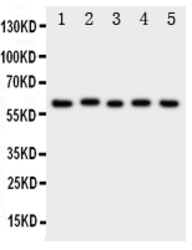
- Additional image