Explore
 Validate
 Learn
Learn Western blot
 Immunocytochemistry
Antibody data
- Antibody Data
- Antigen structure
- References [4]
- Comments [0]
- Validations
- Western blot [2]
- Immunocytochemistry [1]
- Immunohistochemistry [1]
Submit
Validation data
Reference
Comment
Report error
- Product number
- HPA006801 - Provider product page

- Provider
- Atlas Antibodies
- Proper citation
- Atlas Antibodies Cat#HPA006801, RRID:AB_1858899
- Product name
- Anti-XRCC4
- Antibody type
- Polyclonal
- Description
- Polyclonal Antibody against Human XRCC4, Gene description: X-ray repair complementing defective repair in Chinese hamster cells 4, Validated applications: ICC, IHC, WB, Uniprot ID: Q13426, Storage: Store at +4°C for short term storage. Long time storage is recommended at -20°C.
- Reactivity
- Human
- Host
- Rabbit
- Conjugate
- Unconjugated
- Isotype
- IgG
- Vial size
- 100 µl
- Concentration
- 0.2 mg/ml
- Storage
- Store at +4°C for short term storage. Long time storage is recommended at -20°C.
- Handling
- The antibody solution should be gently mixed before use.
Submitted references DNA-PK is activated by SIRT2 deacetylation to promote DNA double-strand break repair by non-homologous end joining
Cell resistance to the Cytolethal Distending Toxin involves an association of DNA repair mechanisms
Organization and dynamics of the nonhomologous end-joining machinery during DNA double-strand break repair
Immunofluorescence and fluorescent-protein tagging show high correlation for protein localization in mammalian cells
Head P, Kapoor-Vazirani P, Nagaraju G, Zhang H, Rath S, Luong N, Haji-Seyed-Javadi R, Sesay F, Wang S, Duong D, Daddacha W, Minten E, Song B, Danelia D, Liu X, Li S, Ortlund E, Seyfried N, Smalley D, Wang Y, Deng X, Dynan W, El-Rayes B, Davis A, Yu D
Nucleic Acids Research 2023;51(15):7972-7987
Nucleic Acids Research 2023;51(15):7972-7987
Cell resistance to the Cytolethal Distending Toxin involves an association of DNA repair mechanisms
Bezine E, Malaisé Y, Loeuillet A, Chevalier M, Boutet-Robinet E, Salles B, Mirey G, Vignard J
Scientific Reports 2016;6(1)
Scientific Reports 2016;6(1)
Organization and dynamics of the nonhomologous end-joining machinery during DNA double-strand break repair
Reid D, Keegan S, Leo-Macias A, Watanabe G, Strande N, Chang H, Oksuz B, Fenyo D, Lieber M, Ramsden D, Rothenberg E
Proceedings of the National Academy of Sciences 2015;112(20)
Proceedings of the National Academy of Sciences 2015;112(20)
Immunofluorescence and fluorescent-protein tagging show high correlation for protein localization in mammalian cells
Stadler C, Rexhepaj E, Singan V, Murphy R, Pepperkok R, Uhlén M, Simpson J, Lundberg E
Nature Methods 2013;10(4):315-323
Nature Methods 2013;10(4):315-323
No comments: Submit comment
Enhanced validation
Enhanced validation
- Submitted by
- klas2
- Enhanced method
- Genetic validation
- Main image
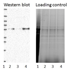
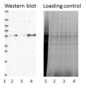
- Experimental details
- Western blot of cell lysate from U-2 OS cells transfected with either siRNA targeting XRCC4 or control siRNA. Lane 1: Marker (250, 130, 95, 72, 55, 36, 28, 17, 10) Lane 2: Cell lysate from U-2OS cells transfected with siRNA targeting XRCC4 Lane 3: N/A Lane 4: Cell lysate from U-2OS cells transfected with control siRNA Right image, lane 1-4: loading control
- Sample type
- U-2 OS
- Primary Ab dilution
- 1:142
- Conjugate
- Horseradish Peroxidase
- Secondary Ab
- Secondary Ab
- Secondary Ab dilution
- 1:3000
- Knockdown/Genetic Approaches Application
- Western blot
Enhanced validation
- Submitted by
- Atlas Antibodies (provider)
- Enhanced method
- Genetic validation
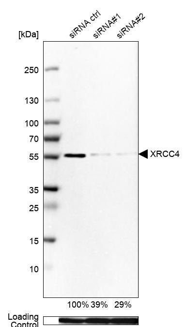
- Main image
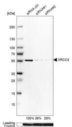
- Experimental details
- Western blot analysis in Rh30 cells transfected with control siRNA, target specific siRNA probe #1 and #2, using Anti-XRCC4 antibody. Remaining relative intensity is presented. Loading control: Anti-GAPDH.
- Sample type
- Human
- Protocol
- Protocol
Supportive validation
- Submitted by
- Atlas Antibodies (provider)
- Main image
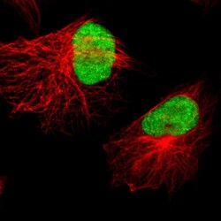
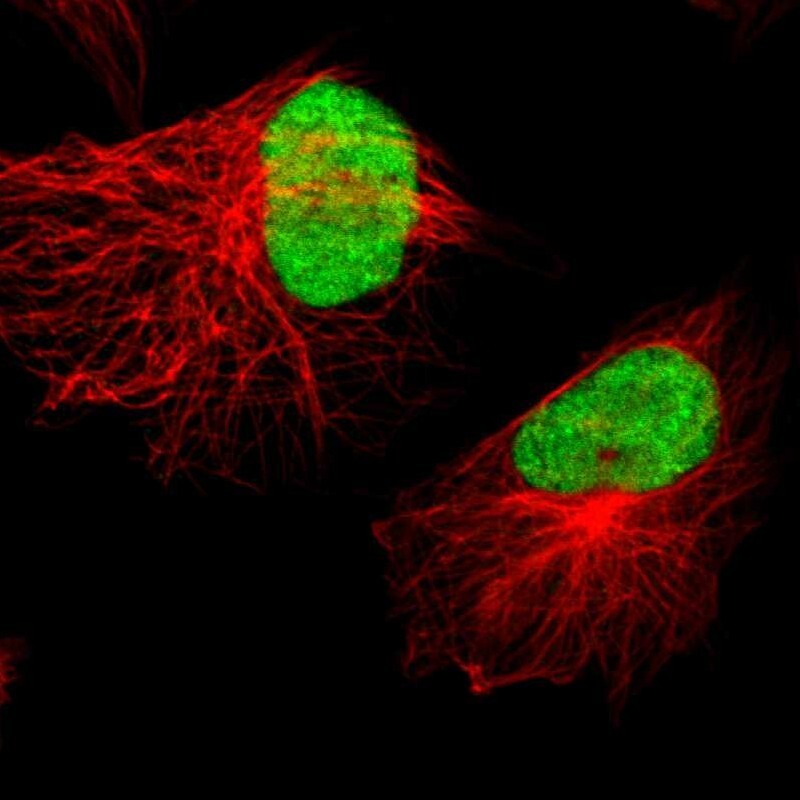
- Experimental details
- Immunofluorescent staining of human cell line U-251 MG shows localization to nucleus.
- Sample type
- Human
Supportive validation
- Submitted by
- Atlas Antibodies (provider)
- Enhanced method
- Orthogonal validation
- Main image

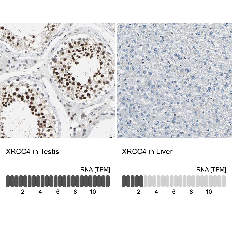
- Experimental details
- Immunohistochemistry analysis in human testis and liver tissues using HPA006801 antibody. Corresponding XRCC4 RNA-seq data are presented for the same tissues.
- Sample type
- Human
- Protocol
- Protocol