 Explore
 Validate
 Learn
Learn Western blot
Antibody data
- Antibody Data
- Antigen structure
- References [0]
- Comments [0]
- Validations
- Western blot [4]
- Immunocytochemistry [5]
- Immunoprecipitation [1]
- Immunohistochemistry [1]
- Other assay [1]
Submit
Validation data
Reference
Comment
Report error
- Product number
- PA5-27518 - Provider product page

- Provider
- Invitrogen Antibodies
- Product name
- FEN1 Polyclonal Antibody
- Antibody type
- Polyclonal
- Antigen
- Recombinant full-length protein
- Description
- Recommended positive controls: HepG2, NIH-3T3, JC, BCL-1, Raw264.7, PC-12. Predicted reactivity: Mouse (96%), Rat (95%), Chicken (81%), Sheep (94%), Rhesus Monkey (98%), Bovine (96%). Store product as a concentrated solution. Centrifuge briefly prior to opening the vial.
- Reactivity
- Human, Mouse, Rat
- Host
- Rabbit
- Isotype
- IgG
- Vial size
- 100 μL
- Concentration
- 0.2 mg/mL
- Storage
- Store at 4°C short term. For long term storage, store at -20°C, avoiding freeze/thaw cycles.
No comments: Submit comment
Supportive validation
- Submitted by
- Invitrogen Antibodies (provider)
- Main image
- Experimental details
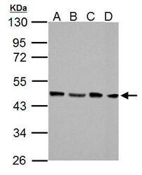
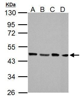
- FEN1 Polyclonal Antibody detects FEN1 protein by western blot analysis. A. 30 µg NIH-3T3 whole cell lysate/extract. B. 30 µg JC whole cell lysate/extract. C. 30 µg BCL-1 whole cell lysate/extract. D. 30 µg Raw264.7 whole cell lysate/extract. 10% SDS-PAGE. FEN1 Polyclonal Antibody (Product # PA5-27518) dilution: 1:1,000. The HRP-conjugated anti-rabbit IgG antibody was used to detect the primary antibody.
- Submitted by
- Invitrogen Antibodies (provider)
- Main image
- Experimental details
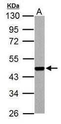
- FEN1 Polyclonal Antibody detects FEN1 protein by western blot analysis. A. 30 µg PC-12 whole cell lysate/extract. 10% SDS-PAGE. FEN1 Polyclonal Antibody (Product # PA5-27518) dilution: 1:1,000. The HRP-conjugated anti-rabbit IgG antibody was used to detect the primary antibody.
- Submitted by
- Invitrogen Antibodies (provider)
- Main image

- Experimental details
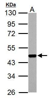
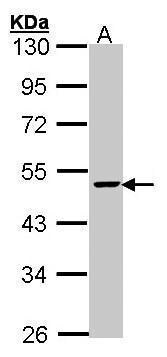
- Western Blot using FEN1 Polyclonal Antibody (Product # PA5-27518). Sample (30 µg of whole cell lysate). Lane A: HepG2. 10% SDS PAGE. FEN1 Polyclonal Antibody (Product # PA5-27518) diluted at 1:1,000. The HRP-conjugated anti-rabbit IgG antibody was used to detect the primary antibody.
- Submitted by
- Invitrogen Antibodies (provider)
- Main image
- Experimental details
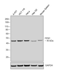
- Western blot was performed using Anti-FEN1 Polyclonal Antibody, (Product # PA5-27518) and a 45 kDa band corresponding to FEN1 was observed in the cell lines and tissue tested. Modified whole cell extracts (1%SDS) (30 µg lysate) of A-431 (Lane 1), HCT 116 (Lane 2), HeLa (Lane 3), Hep G2 (Lane 4) and Mouse Spleen (Lane 5) were electrophoresed using Novex® NuPAGE® 4-12 % Bis-Tris gel (Product # NP0321BOX). Resolved proteins were then transferred onto a nitrocellulose membrane (Product # IB23001) by iBlot® 2 Dry Blotting System (Product # IB21001). The blot was probed with the primary antibody (1:1000 dilution) and detected by chemiluminescence with Goat anti-Rabbit IgG (Heavy Chain), Superclonal™ Recombinant Secondary Antibody, HRP conjugate (Product # A27036, 1:4000 dilution) using the iBright FL 1000 (Product # A32752). Chemiluminescent detection was performed using Novex® ECL Chemiluminescent Substrate Reagent Kit (Product # WP20005)..

Supportive validation
- Submitted by
- Invitrogen Antibodies (provider)
- Main image

- Experimental details
- Immunofluorescent analysis of FEN1 in paraformaldehyde-fixed A549 cells using a FEN1 polyclonal antibody (Product # PA5-27518) at a 1:200 dilution.
- Submitted by
- Invitrogen Antibodies (provider)
- Main image
- Experimental details
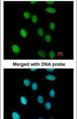
- FEN1 Polyclonal Antibody detects FEN1 protein at nucleus by immunofluorescent analysis. Sample: HeLa cells were fixed in 4% paraformaldehyde at RT for 15 min. Green: FEN1 stained by FEN1 Polyclonal Antibody (Product # PA5-27518) diluted at 1:500. Red: alpha Tubulin, a cytoskeleton marker, stained by alpha Tubulin Polyclonal Antibody [GT114] (Product # MA5-31466) diluted at 1:1,000. Blue: Fluoroshield with DAPI .

- Submitted by
- Invitrogen Antibodies (provider)
- Main image

- Experimental details
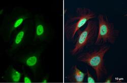
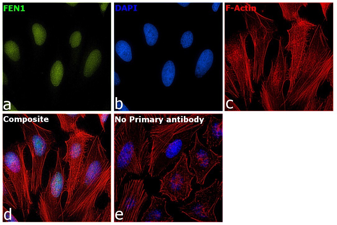
- Immunofluorescence analysis of FEN1 was performed using 70% confluent log phase HeLa cells. The cells were fixed with 4% paraformaldehyde for 10 minutes, permeabilized with 0.1% Triton™ X-100 for 15 minutes, and blocked with 2% BSA for 1 hour at room temperature. The cells were labeled with FEN1 Polyclonal Antibody (Product # PA5-27518) at 1:200 dilution in 0.1% BSA, incubated at 4 degree celsius overnight and then with Goat anti-Rabbit IgG (H+L), Superclonal™ Recombinant Secondary Antibody, Alexa Fluor 488 (Product # A27034) at a dilution of 1:2000 for 45 minutes at room temperature (Panel a: green). Nuclei (Panel b: blue) were stained with SlowFade® Gold Antifade Mountant with DAPI (Product # S36938). F-actin (Panel c: red) was stained with Rhodamine Phalloidin (Product # R415, 1:300). Panel d represents the merged image showing staining in nucleus. Panel e represents control cells with no primary antibody to assess background. The images were captured at 60X magnification.
- Submitted by
- Invitrogen Antibodies (provider)
- Main image

- Experimental details
- FEN1 Polyclonal Antibody detects FEN1 protein at nucleus by immunofluorescent analysis. Sample: HeLa cells were fixed in 4% paraformaldehyde at RT for 15 min. Green: FEN1 stained by FEN1 Polyclonal Antibody (Product # PA5-27518) diluted at 1:500. Red: alpha Tubulin, a cytoskeleton marker, stained by alpha Tubulin Polyclonal Antibody [GT114] (Product # MA5-31466) diluted at 1:1,000. Blue: Fluoroshield with DAPI .

- Submitted by
- Invitrogen Antibodies (provider)
- Main image
- Experimental details
- Immunofluorescence analysis of FEN1 was performed using 70% confluent log phase HeLa cells. The cells were fixed with 4% paraformaldehyde for 10 minutes, permeabilized with 0.1% Triton™ X-100 for 15 minutes, and blocked with 2% BSA for 1 hour at room temperature. The cells were labeled with FEN1 Polyclonal Antibody (Product # PA5-27518) at 1:200 dilution in 0.1% BSA, incubated at 4 degree celsius overnight and then with Goat anti-Rabbit IgG (Heavy Chain), Superclonal™ Recombinant Secondary Antibody, Alexa Fluor 488 (Product # A27034) at a dilution of 1:2000 for 45 minutes at room temperature (Panel a: green). Nuclei (Panel b: blue) were stained with SlowFade® Gold Antifade Mountant with DAPI (Product # S36938). F-actin (Panel c: red) was stained with Rhodamine Phalloidin (Product # R415, 1:300). Panel d represents the merged image showing staining in nucleus. Panel e represents control cells with no primary antibody to assess background. The images were captured at 60X magnification.

Supportive validation
- Submitted by
- Invitrogen Antibodies (provider)
- Main image

- Experimental details
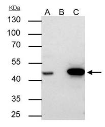
- FEN1 Polyclonal Antibody immunoprecipitates FEN1 protein in IP experiments. IP samples: Jurkat whole cell extract. A. 30 µg Jurkat whole cell extract. B. Control with 4 µg of preimmune Rabbit IgG. C. Immunoprecipitation of FEN1 protein by 4 µg FEN1 Polyclonal Antibody (Product # PA5-27518). 10 % SDS-PAGE. The immunoprecipitated FEN1 protein was detected by FEN1 Polyclonal Antibody (Product # PA5-27518) diluted at 1:500.

Supportive validation
- Submitted by
- Invitrogen Antibodies (provider)
- Main image
- Experimental details
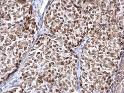
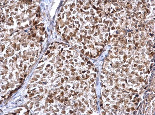
- FEN1 Polyclonal Antibody detects FEN1 protein at nucleus on human breast carcinoma by immunohistochemical analysis. Sample: Paraffin-embedded human breast carcinoma. FEN1 Polyclonal Antibody (Product # PA5-27518) diluted at 1:500. Antigen Retrieval: EDTA based buffer, pH 8.0, 15 min.
Supportive validation
- Submitted by
- Invitrogen Antibodies (provider)
- Main image
- Experimental details
- FEN1 Polyclonal Antibody immunoprecipitates FEN1 protein in IP experiments. IP samples: Jurkat whole cell extract. A. 30 µg Jurkat whole cell extract. B. Control with 4 µg of preimmune Rabbit IgG. C. Immunoprecipitation of FEN1 protein by 4 µg FEN1 Polyclonal Antibody (Product # PA5-27518). 10 % SDS-PAGE. The immunoprecipitated FEN1 protein was detected by FEN1 Polyclonal Antibody (Product # PA5-27518) diluted at 1:500.
