Explore
 Validate
 Learn
Learn Western blot
 ELISA
ELISAAntibody data
- Antibody Data
- Antigen structure
- References [0]
- Comments [0]
- Validations
- Western blot [2]
- ELISA [4]
Submit
Validation data
Reference
Comment
Report error
- Product number
- LS-C829079 - Provider product page

- Provider
- LSBio
- Product name
- POU2F3 / PLA-1 Antibody (clone 34H8) LS-C829079
- Antibody type
- Monoclonal
- Description
- Protein A purified
- Reactivity
- Mouse
- Host
- Mouse
- Isotype
- IgG
- Antibody clone number
- 34H8
- Storage
- Store lyophilized at -20°C for up to 1 year. Store reconstituted at 2°C to 8°C for up to 3 weeks. Aliquot and store at -20°C or below for up to 1 year. Avoid freeze/thaw cycles.
No comments: Submit comment
Supportive validation
- Submitted by
- LSBio (provider)
- Main image
- Experimental details
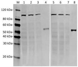
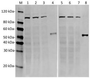
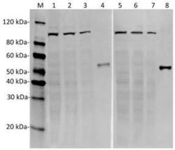
- Western Blot of Sp2/0 cell lysates in different concentration with two independent antibodies: Mouse Epoc-1 Antibody (34H8) and Mouse Epoc-1 Antibody (39F11). The correlated pattern indicates the high specificity of these two antibodies. The different concentration of cell lysates indicates the high affinity and sensitivity of the antibodies. Lane 1: 100 µg Sp2/0 cell lysate. Lane 2: 50 µg Sp2/0 cell lysate. Lane 3: 25 µg Sp2/0 cell lysate. Lane 4: 50 ng recombinant mouse Epoc-1 protein. Lane 5: 100 µg Sp2/0 cell lysate. Lane 6: 50 µg Sp2/0 cell lysate. Lane 7: 25 µg Sp2/0 cell lysate. Lane 8: 50 ng recombinant mouse Epoc-1 protein. Primary Antibody: Lane 1~4: Mouse Epoc-1 Antibody (34H8) 1 µg/ml. Lane 5~8: Mouse Epoc-1 Antibody (39F11) 1 µg/ml. Secondary Antibody: Goat anti-Mouse IgG (H&L) [IRDye800] 0.125 µg/ml.
- Submitted by
- LSBio (provider)
- Main image
- Experimental details
- Western Blot of Sp2/0 cell lysates in different concentration with two independent antibodies: Mouse Epoc-1 Antibody (34H8) and Mouse Epoc-1 Antibody (39F11). The correlated pattern indicates the high specificity of these two antibodies. The different concentration of cell lysates indicates the high affinity and sensitivity of the antibodies. Lane 1: 100 µg Sp2/0 cell lysate. Lane 2: 50 µg Sp2/0 cell lysate. Lane 3: 25 µg Sp2/0 cell lysate. Lane 4: 50 ng recombinant mouse Epoc-1 protein. Lane 5: 100 µg Sp2/0 cell lysate. Lane 6: 50 µg Sp2/0 cell lysate. Lane 7: 25 µg Sp2/0 cell lysate. Lane 8: 50 ng recombinant mouse Epoc-1 protein. Primary Antibody: Lane 1~4: Mouse Epoc-1 Antibody (34H8) 1 µg/ml. Lane 5~8: Mouse Epoc-1 Antibody (39F11) 1 µg/ml. Secondary Antibody: Goat anti-Mouse IgG (H&L) [IRDye800] 0.125 µg/ml.

Supportive validation
- Submitted by
- LSBio (provider)
- Enhanced method
- Genetic validation
- Main image
- Experimental details
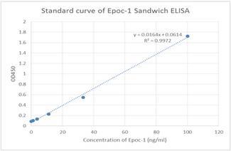
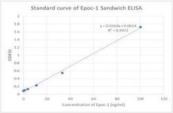
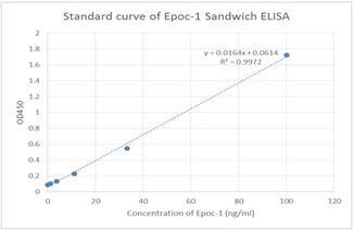
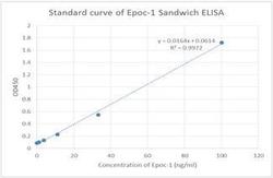
- Standard curve of Epoc-1 Sandwich ELISA. The Epoc-1 Sandwich ELISA assay is developed by using Mouse Epoc-1 Antibody (34H8) and Mouse Epoc-1 Antibody (39F11) as capture and detection antibody, respectively. These two antibodies recognize different epitopes. In this ELISA assay, Mouse Epoc-1 Antibody (39F11) was labeled with Biotin. The sensitivity is < 1 ng/ml and the detection range is 0-100 ng/ml.
- Submitted by
- LSBio (provider)
- Enhanced method
- Genetic validation
- Main image
- Experimental details
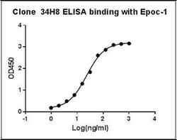
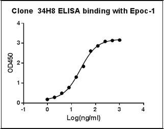
- ELISA binding of Mouse Epoc-1 Antibody (34H8) with recombinant Mouse Epoc-1 protein. Coating antigen: Epoc-1, 1 ug/ml. Epoc-1 antibody dilution start from 1000 ng/ml, EC50= 23.12 ng/ml.

- Submitted by
- LSBio (provider)
- Main image

- Experimental details
- ELISA binding of Mouse Epoc-1 Antibody (34H8) with recombinant Mouse Epoc-1 protein. Coating antigen: Epoc-1, 1 ug/ml. Epoc-1 antibody dilution start from 1000 ng/ml, EC50= 23.12 ng/ml.
- Submitted by
- LSBio (provider)
- Main image
- Experimental details
- Standard curve of Epoc-1 Sandwich ELISA. The Epoc-1 Sandwich ELISA assay is developed by using Mouse Epoc-1 Antibody (34H8) and Mouse Epoc-1 Antibody (39F11) as capture and detection antibody, respectively. These two antibodies recognize different epitopes. In this ELISA assay, Mouse Epoc-1 Antibody (39F11) was labeled with Biotin. The sensitivity is < 1 ng/ml and the detection range is 0-100 ng/ml.