 Explore
 Validate
 Learn
Learn Western blot
Antibody data
- Antibody Data
- Antigen structure
- References [3]
- Comments [0]
- Validations
- Western blot [1]
- Immunohistochemistry [1]
- Flow cytometry [1]
Submit
Validation data
Reference
Comment
Report error
- Product number
- AP8161a - Provider product page

- Provider
- Abcepta
- Proper citation
- Abgent Cat#AP8161a, RRID:AB_2165182
- Product name
- Urokinase (PLAU) Antibody (N-term)
- Antibody type
- Polyclonal
- Antigen
- Synthetic peptide
- Description
- Purified Rabbit Polyclonal Antibody (Pab)
- Reactivity
- Human, Mouse
- Host
- Rabbit
- Isotype
- IgG
- Vial size
- 400 µl
- Concentration
- 2 mg/ml
- Storage
- Maintain refrigerated at 2-8°C for up to 6 months. For long term storage store at -20°C in small aliquots to prevent freeze-thaw cycles.
Submitted references Suppression of tumor growth in H-ras12V liver cancer mice by delivery of programmed cell death protein 4 using galactosylated poly(ethylene glycol)-chitosan-graft-spermine.
In vivo suppression of vein graft disease by nonviral, electroporation-mediated, gene transfer of tissue inhibitor of metalloproteinase-1 linked to the amino terminal fragment of urokinase (TIMP-1.ATF), a cell-surface directed matrix metalloproteinase inhibitor.
A gene expression signature that distinguishes desmoid tumours from nodular fasciitis.
Kim JH, Minai-Tehrani A, Kim YK, Shin JY, Hong SH, Kim HJ, Lee HD, Chang SH, Yu KN, Bang YB, Cho CS, Yoon TJ, Yu DY, Jiang HL, Cho MH
Biomaterials 2012 Feb;33(6):1894-902
Biomaterials 2012 Feb;33(6):1894-902
In vivo suppression of vein graft disease by nonviral, electroporation-mediated, gene transfer of tissue inhibitor of metalloproteinase-1 linked to the amino terminal fragment of urokinase (TIMP-1.ATF), a cell-surface directed matrix metalloproteinase inhibitor.
Eefting D, de Vries MR, Grimbergen JM, Karper JC, van Bockel JH, Quax PH
Journal of vascular surgery 2010 Feb;51(2):429-37
Journal of vascular surgery 2010 Feb;51(2):429-37
A gene expression signature that distinguishes desmoid tumours from nodular fasciitis.
Bacac M, Migliavacca E, Stehle JC, McKee T, Delorenzi M, Coindre JM, Guillou L, Stamenkovic I
The Journal of pathology 2006 Mar;208(4):543-53
The Journal of pathology 2006 Mar;208(4):543-53
No comments: Submit comment
Supportive validation
- Submitted by
- Abcepta (provider)
- Main image
- Experimental details
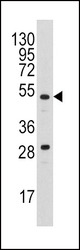
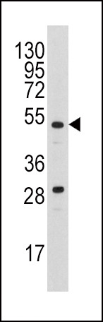
- Western blot analysis of anti-PLAU Antibody (N-term) (Cat.#AP8161a) in A2058 cell line lysates (35ug/lane). PLAU (arrow) was detected using the purified Pab.
- Primary Ab dilution
- 1:1000
Supportive validation
- Submitted by
- Abcepta (provider)
- Main image
- Experimental details
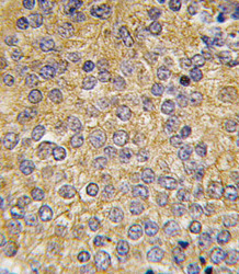
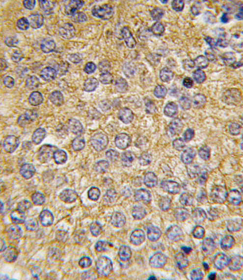
- "Formalin-fixed and paraffin-embedded human prostata carcinoma tissue reacted with PLAU antibody (N-term), which was peroxidase-conjugated to the secondary antibody, followed by DAB staining. This data demonstrates the use of this antibody for immunohistochemistry; clinical relevance has not been evaluated."
- Primary Ab dilution
- 1:10~50
Supportive validation
- Submitted by
- Abcepta (provider)
- Main image
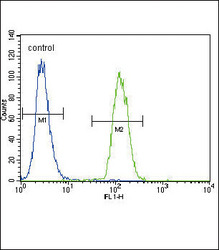
- Experimental details
- Urokinase (PLAU) Antibody (N-term) (Cat. #AP8161a) flow cytometric analysis of A2058 cells (right histogram) compared to a negative control cell (left histogram).FITC-conjugated goat-anti-rabbit secondary antibodies were used for the analysis.
- Primary Ab dilution
- 1:10~50
