 Explore
 Validate
 Learn
Learn Western blot
 Immunocytochemistry
Antibody data
- Antibody Data
- Antigen structure
- References [1]
- Comments [0]
- Validations
- Immunocytochemistry [2]
- Other assay [1]
Submit
Validation data
Reference
Comment
Report error
- Product number
- PA5-18822 - Provider product page

- Provider
- Invitrogen Antibodies
- Product name
- 14-3-3 theta Polyclonal Antibody
- Antibody type
- Polyclonal
- Antigen
- Synthetic peptide
- Description
- This antibody is predicted to react with canine, mouse and rat based on sequence homology. This antibody is tested in Peptide ELISA: antibody detection limit dilution 4,000.
- Reactivity
- Human
- Host
- Goat
- Isotype
- IgG
- Vial size
- 100 μg
- Concentration
- 0.5 mg/mL
- Storage
- -20°C, Avoid Freeze/Thaw Cycles
Submitted references Prospective evaluation of the lymph node proteome in dogs with multicentric lymphoma supplemented with sulforaphane.
Parachini-Winter C, Bracha S, Ramsey SA, Yang L, Ho E, Leeper HJ, Curran KM
Journal of veterinary internal medicine 2020 Sep;34(5):2036-2047
Journal of veterinary internal medicine 2020 Sep;34(5):2036-2047
No comments: Submit comment
Supportive validation
- Submitted by
- Invitrogen Antibodies (provider)
- Main image
- Experimental details
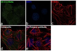
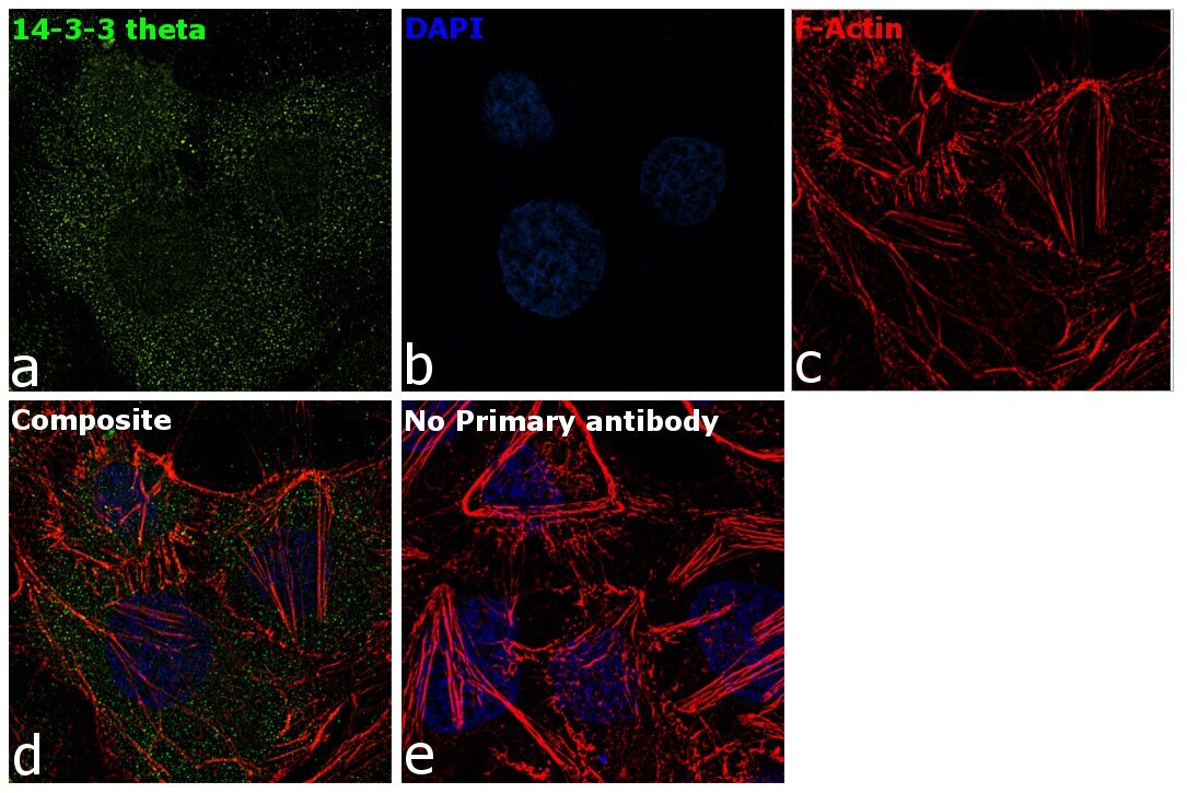
- Immunofluorescence analysis of 14-3-3 theta was performed using HeLa cells. The cells were fixed with 4% paraformaldehyde for 10 minutes, permeabilized with 0.1% Triton™ X-100 for 15 minutes, and blocked with 2% BSA for 1 hour at room temperature. The cells were labeled with 14-3-3 theta Goat Polyclonal Antibody (Product # PA5-18822) at 5 µg/mL in 0.1% BSA and incubated overnight at 4 degree and then labeled with Rabbit anti-Goat IgG (H+L) Cross-Adsorbed Secondary Antibody, Alexa Fluor 488 (Product # A-11078) at a dilution of 1:2000 for 45 minutes at room temperature (Panel a: green). Nuclei (Panel b: blue) were stained with ProLong™ Diamond Antifade Mountant with DAPI (Product # P36962). F-actin (Panel c: red) was stained with Rhodamine Phalloidin (Product # R415, 1:300). Panel d represents the composite image showing cytoplasmic localization of 14-3-3 theta in HeLa cells . Panel e represents control cells with no primary antibody to assess background. The images were captured at 60X magnification..
- Submitted by
- Invitrogen Antibodies (provider)
- Main image

- Experimental details
- Immunofluorescence analysis of 14-3-3 theta was performed using HeLa cells. The cells were fixed with 4% paraformaldehyde for 10 minutes, permeabilized with 0.1% Triton™ X-100 for 15 minutes, and blocked with 2% BSA for 1 hour at room temperature. The cells were labeled with 14-3-3 theta Goat Polyclonal Antibody (Product # PA5-18822) at 5 µg/mL in 0.1% BSA and incubated overnight at 4 degree and then labeled with Rabbit anti-Goat IgG (H+L) Cross-Adsorbed Secondary Antibody, Alexa Fluor 488 (Product # A-11078) at a dilution of 1:2000 for 45 minutes at room temperature (Panel a: green). Nuclei (Panel b: blue) were stained with ProLong™ Diamond Antifade Mountant with DAPI (Product # P36962). F-actin (Panel c: red) was stained with Rhodamine Phalloidin (Product # R415, 1:300). Panel d represents the composite image showing cytoplasmic localization of 14-3-3 theta in HeLa cells . Panel e represents control cells with no primary antibody to assess background. The images were captured at 60X magnification..

Supportive validation
- Submitted by
- Invitrogen Antibodies (provider)
- Main image
- Experimental details
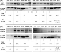
- FIGURE 2 Immunoblots of lymph node samples for 7 dogs (d) with naive multicentric lymphoma before (D0) and after (D7) supplementation with sulforaphane. The proteins HIP, A, and 14-3-3-theta, B, were detected in all samples (top rows). The immunoblots for beta-actin (second rows) were used to normalize the band volume of HIP and 14-3-3-theta. The normalized expression ratio at D7/D0 is the ratio of the normalized band volume at D7 vs D0 for each dog (third rows). For comparison, the corresponding fold change at D7 vs D0 previously determined by LC-MS/MS is also annotated (bottom rows). FC D7/D0: Fold change at D7 vs D0 (mass spectrometry); NA: Not applicable; NER D7/D0: Normalized expression ratio at D7 vs D0 (immunoblot)
