 Explore
 Validate
 Learn
Learn Western blot
 ELISA
ELISAAntibody data
- Antibody Data
- Antigen structure
- References [0]
- Comments [0]
- Validations
- Western blot [1]
- Immunohistochemistry [4]
Submit
Validation data
Reference
Comment
Report error
- Product number
- LS-C745337 - Provider product page

- Provider
- LSBio
- Product name
- BLIMP1 / PRDM1 Antibody (C-Terminus) LS-C745337
- Antibody type
- Polyclonal
- Description
- Affinity purified
- Reactivity
- Human, Mouse, Rat, Canine
- Host
- Rabbit
- Isotype
- IgG
- Storage
- Store vial at -20°C or below prior to opening. Dilute 1:10 to minimize loss. Store the vial at -20°C or below after dilution. Avoid freeze-thaw cycles.
No comments: Submit comment
Enhanced validation
- Submitted by
- LSBio (provider)
- Enhanced method
- Genetic validation
- Main image

- Experimental details
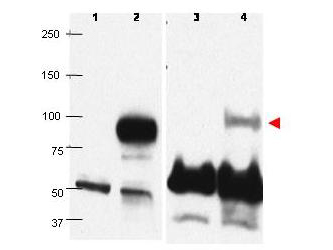
- Western blots using affinity purified anti-PRDM1/BLIMP1 antibody show detection of overexpressed PRDM1/BLIMP1 in whole transfected Raji cell lysate (lane 2) at ~88kDa. Lane 1 shows mock transfection in whole Raji cell lysate. Detection of endogenous PRDM1/BLIMP1 (lane 4) is illustrated in human plasma cell nuclear extract, but not in Raji whole cell nuclear extract (lane 3). The identity of the lower dark band at ~50-60kDa is unknown. Primary antibody was used at a 1:1000 dilution in 5% PBS-Tween.
Enhanced validation
- Submitted by
- LSBio (provider)
- Enhanced method
- Genetic validation
- Main image
- Experimental details
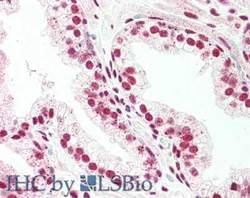
- Immunohistochemistry of rabbit anti-PRDM1-BLMP1 antibody. Tissue: prostate. Fixation: formalin fixed paraffin embedded. Antigen retrieval: not required. Primary antibody: Anti-PRDM1-BLMP1 at 5 µg/mL for 1 h at RT. Secondary antibody: Peroxidase rabbit secondary antibody at 1:10,000 for 45 min at RT. Staining: PRDM1-BLMP1 as precipitated red signal with hematoxylin purple nuclear counterstain.

- Submitted by
- LSBio (provider)
- Enhanced method
- Genetic validation
- Main image

- Experimental details
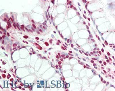
- Immunohistochemistry of rabbit anti-PRDM1-BLMP1 antibody. Tissue: colon. Fixation: formalin fixed paraffin embedded. Antigen retrieval: not required. Primary antibody: Anti-PRDM1-BLMP1 at 5 µg/mL for 1 h at RT. Secondary antibody: Peroxidase rabbit secondary antibody at 1:10,000 for 45 min at RT. Staining: PRDM1-BLMP1 as precipitated red signal with hematoxylin purple nuclear counterstain.

- Submitted by
- LSBio (provider)
- Main image

- Experimental details
- Immunohistochemistry of rabbit anti-PRDM1-BLMP1 antibody. Tissue: colon. Fixation: formalin fixed paraffin embedded. Antigen retrieval: not required. Primary antibody: Anti-PRDM1-BLMP1 at 5 µg/mL for 1 h at RT. Secondary antibody: Peroxidase rabbit secondary antibody at 1:10,000 for 45 min at RT. Staining: PRDM1-BLMP1 as precipitated red signal with hematoxylin purple nuclear counterstain.
- Submitted by
- LSBio (provider)
- Main image

- Experimental details
- Immunohistochemistry of rabbit anti-PRDM1-BLMP1 antibody. Tissue: prostate. Fixation: formalin fixed paraffin embedded. Antigen retrieval: not required. Primary antibody: Anti-PRDM1-BLMP1 at 5 µg/mL for 1 h at RT. Secondary antibody: Peroxidase rabbit secondary antibody at 1:10,000 for 45 min at RT. Staining: PRDM1-BLMP1 as precipitated red signal with hematoxylin purple nuclear counterstain.
