Explore
 Validate
 Learn
Learn Western blot
 Immunocytochemistry
Antibody data
- Antibody Data
- Antigen structure
- References [1]
- Comments [0]
- Validations
- Immunocytochemistry [1]
- Immunohistochemistry [3]
- Flow cytometry [2]
- Other assay [1]
Submit
Validation data
Reference
Comment
Report error
- Product number
- PA5-19216 - Provider product page

- Provider
- Invitrogen Antibodies
- Product name
- Blimp-1 Polyclonal Antibody
- Antibody type
- Polyclonal
- Antigen
- Synthetic peptide
- Description
- This antibody is predicted to react with bovine, canine, mouse, porcine and rat based on sequence homology. This antibody is tested in Peptide ELISA: antibody detection limit dilution 1,000.
- Reactivity
- Human
- Host
- Goat
- Isotype
- IgG
- Vial size
- 100 μg
- Concentration
- 0.5 mg/mL
- Storage
- -20°C, Avoid Freeze/Thaw Cycles
Submitted references A Novel Role for the Regulatory Nod-Like Receptor NLRP12 in Anti-Dengue Virus Response.
Li X, Dong Z, Liu Y, Song W, Pu J, Jiang G, Wu Y, Liu L, Huang X
Frontiers in immunology 2021;12:744880
Frontiers in immunology 2021;12:744880
No comments: Submit comment
Supportive validation
- Submitted by
- Invitrogen Antibodies (provider)
- Main image
- Experimental details
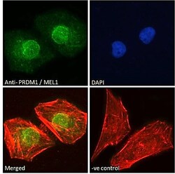
- Immunocytochemistry analysis of Blimp-1 using Blimp-1 Polyclonal Antibody (Product # PA5-19216) in paraformaldehyde fixed HeLa cells, permeabilized with 0.15% Triton. Primary incubation 1hr (10 µg/mL) followed by Alexa Fluor 488 secondary antibody (2 µg/mL), showing nuclear staining. Actin filaments were stained with phalloidin (red) and the nuclear stain is DAPI (blue). Negative control: Unimmunized goat IgG (10 µg/mL) followed by Alexa Fluor 488 secondary antibody (2 µg/mL).

Supportive validation
- Submitted by
- Invitrogen Antibodies (provider)
- Main image
- Experimental details
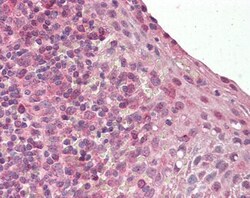
- Immunohistochemistry analysis of Blimp-1 in human tonsil. Samples were incubated with Blimp-1 polyclonal antibody (Product # PA5-19216) using a dilution of 3.75 µg/mL. Formalin-fixed, paraffin-embedded tissue after heat-induced antigen retrieval.
- Submitted by
- Invitrogen Antibodies (provider)
- Main image
- Experimental details
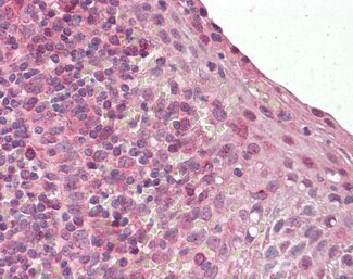
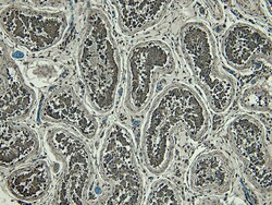
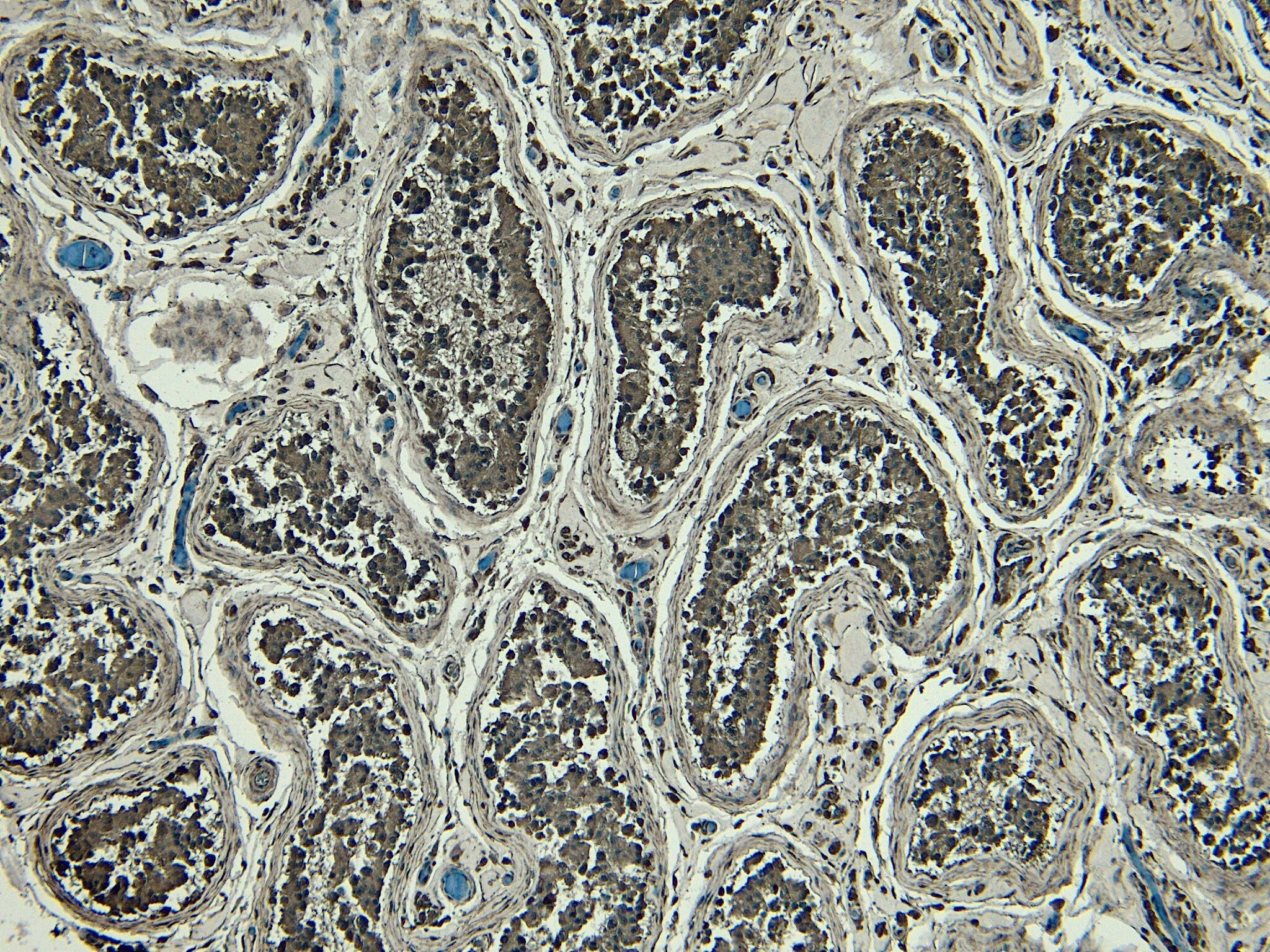
- Immunohistochemistry (PFA fixed) analysis of Blimp-1 using Blimp-1 Polyclonal Antibody (Product # PA5-19216) (6 µg/mL) in staining of paraffin embedded Human Testis. Heat induced antigen retrieval with citrate buffer pH 6, HRP-staining.
- Submitted by
- Invitrogen Antibodies (provider)
- Main image
- Experimental details
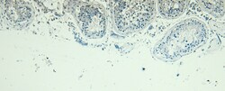
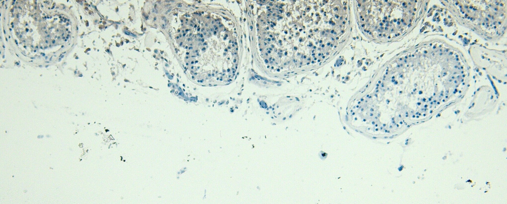
- Negative Control for Blimp-1 Polyclonal Antibody (Product # PA5-19216), showing immunohistochemical staining of paraffin embedded Human Testis, with no primary antibody.
Supportive validation
- Submitted by
- Invitrogen Antibodies (provider)
- Main image
- Experimental details
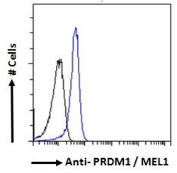
- Flow cytometric analysis of Blimp-1 in A431 cells using a polyclonal antibody (Product #PA5-19216). A431 cells (blue line) were paraformaldehyde fixed and permeabilized with 0.5% Triton. The primary antibody was incubated for one hour (10 µg/mL) followed by an Alexa Fluor 488 secondary antibody (1 µg/mL). IgG control: Unimmunized goat IgG (black line) followed by an Alexa Fluor 488 secondary antibody.
- Submitted by
- Invitrogen Antibodies (provider)
- Main image

- Experimental details
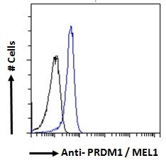
- Flow cytometric analysis of Blimp-1 in A431 cells using a polyclonal antibody (Product #PA5-19216). A431 cells (blue line) were paraformaldehyde fixed and permeabilized with 0.5% Triton. The primary antibody was incubated for one hour (10 µg/mL) followed by an Alexa Fluor 488 secondary antibody (1 µg/mL). IgG control: Unimmunized goat IgG (black line) followed by an Alexa Fluor 488 secondary antibody.

Supportive validation
- Submitted by
- Invitrogen Antibodies (provider)
- Main image
- Experimental details
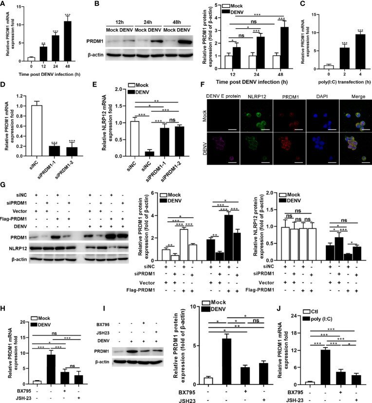
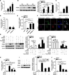
- Figure 2 DENV suppressed NLRP12 expression by up-regulating PRDM-1 expression in a manner dependent on the TBK1/IRF3 and NF-kappaB signaling pathways. (A, B) hMDMs were infected with DENV (MOI=4) for indicated time periods and PRDM1 expression was determined by qPCR and western blot. The gray value of PRDM1 protein bands was analyzed by Image J gel image analysis software, and the relative grayscale value was normalized to that of beta-actin. (C) hMDMs were treated with Poly ( I:C ) and harvested for PRDM1 expression analysis. (D) The siRNA knockdown of PRDM1 was performed in hMDMs and the mRNA level of PRDM1 was measured. (E) PRDM1-silenced hMDMs were infected with DENV for 48h (MOI=4) and NLRP12 expression was determined via qPCR. (F) dTHP1 cells were infected with DENV with an MOI of 4 for 72h. Immunofluorescence staining was performed for DENV E protein, NLRP12, PRDM1 and DAPI (Bar: 10mum). (G) dTHP1 cells were transiently transfected with the siRNAs against the PRDM1, and then PRDM1-Flag-expressing vectors were transfected into the cells. After DENV infection at a MOI of 4 for 72h, the expression levels of NLRP12 and PRDM1 were detected by western blot analysis. The gray value of PRDM1 protein and NLRP12 bands was analyzed by Image J gel image analysis software and the relative grayscale value was normalized to that of beta-actin. (H, I) hMDMs cells were pre-treated with the inhibitors for TBK1 (BX795) or NF-kappaB (JSH-23) and subjected to DENV infection at a MOI of 4 f