Explore
 Validate
 Learn
LearnPA5-78713
antibody from Invitrogen Antibodies
Targeting: ACSL1
ACS1, FACL1, FACL2, LACS, LACS1, LACS2
 Western blot
Antibody data
- Antibody Data
- Antigen structure
- References [0]
- Comments [0]
- Validations
- Western blot [3]
- Immunocytochemistry [2]
- Immunohistochemistry [3]
- Flow cytometry [1]
Submit
Validation data
Reference
Comment
Report error
- Product number
- PA5-78713 - Provider product page

- Provider
- Invitrogen Antibodies
- Product name
- ACSL1 Polyclonal Antibody
- Antibody type
- Polyclonal
- Antigen
- Recombinant full-length protein
- Description
- Reconstitute with 0.2 mL of distilled water to yield a concentration of 500 µg/mL.
- Reactivity
- Human, Mouse, Rat
- Host
- Rabbit
- Isotype
- IgG
- Vial size
- 100 µg
- Concentration
- 500 µg/mL
- Storage
- -20°C
No comments: Submit comment
Supportive validation
- Submitted by
- Invitrogen Antibodies (provider)
- Main image
- Experimental details
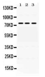
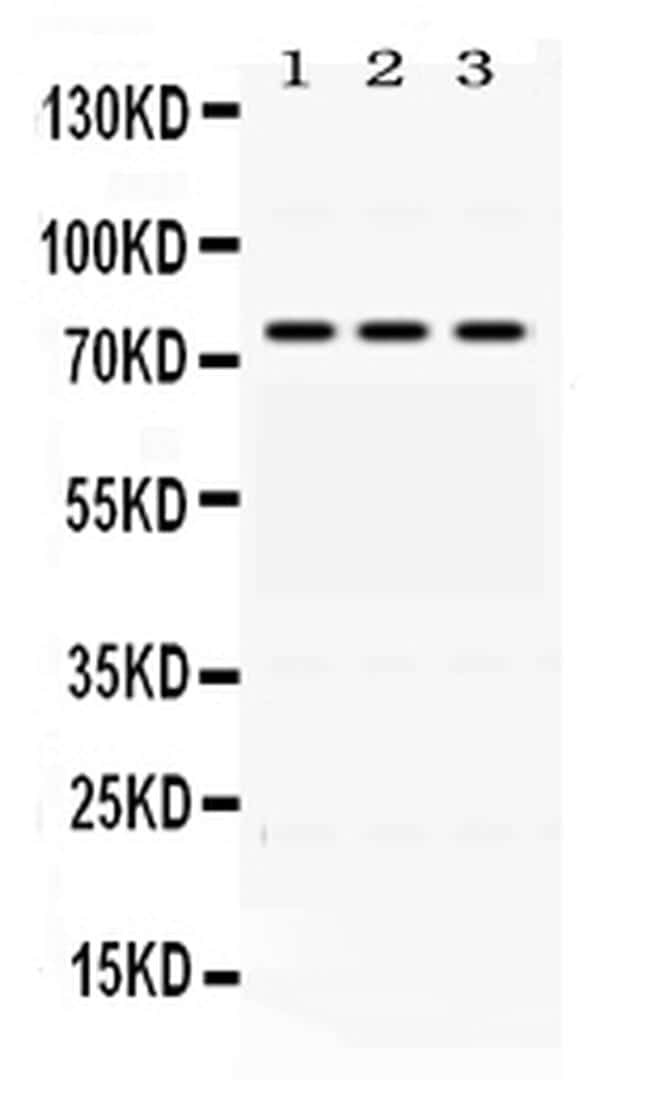
- Western blot analysis of ACSL1 in rat liver extract (lane 1) HEPA whole cell lysate (lane 2) and A549 whole cell lysate (lane 3). Sample was incubated with ACSL1 polyclonal antibody (Product # PA5-78713) at a dilution of 0.5 µg/mL. Signal development was performed using a chemiluminescence (ECL) kit.
- Submitted by
- Invitrogen Antibodies (provider)
- Main image
- Experimental details
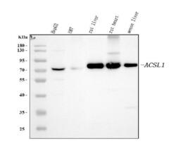
- Western blot analysis of ACSL1 in, Lane 1: human HepG2 whole cell lysates, Lane 2: human U87 whole cell lysates, Lane 3: rat liver tissue lysates, Lane 4: rat heart tissue lysates, Lane 5: mouse liver tissue lysates. Electrophoresis was performed on a 5-20% SDS-PAGE gel at 70V (Stacking gel) / 90V (Resolving gel) for 2-3 hours. The sample well of each lane was loaded with 30 µg of sample under reducing conditions. After Electrophoresis, proteins were transferred to a nitrocellulose membrane at 150 mA for 50-90 minutes. The membrane was blocked with 5% non-fat milk/TBS for 1. 5 hour at RT. The membrane was incubated with ACSL1 Polyclonal Antibody (Product # PA5-78713) at 0.5 μg/mL overnight at 4°C, then washed with TBS-0. 1% Tween 3 times with 5 minutes each and probed with a goat anti-rabbit IgG-HRP secondary antibody at a dilution of 1:5,000 for 1. 5 hour at RT. The signal is developed using an Enhanced Chemiluminescent detection (ECL) kit. A specific band was detected for ACSL1 at approximately 78 kDa. The expected band size for ACSL1 is at 78 kDa.

- Submitted by
- Invitrogen Antibodies (provider)
- Main image
- Experimental details
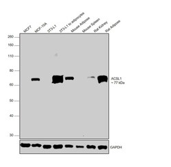
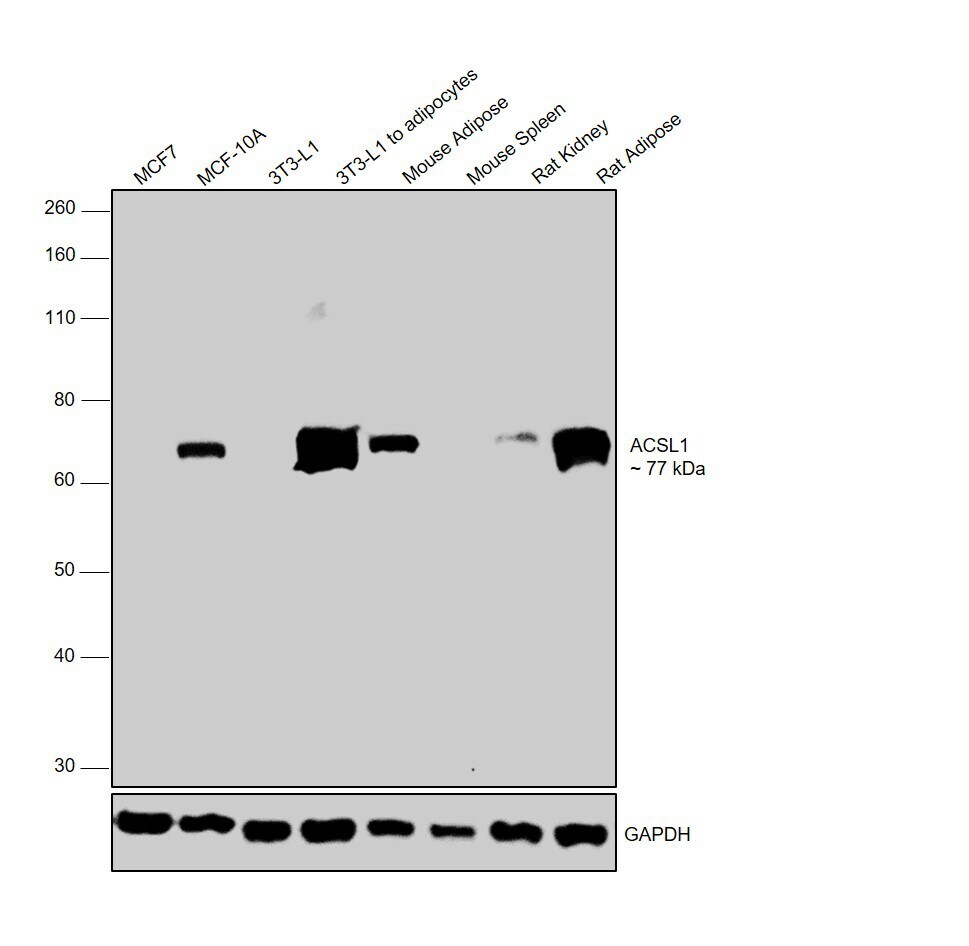
- Western blot was performed using Anti-ACSL1 Polyclonal Antibody (Product # PA5-78713) and a 77 kDa band corresponding to ACSL1 was observed across cell lines and tissues tested except in MCF7, undifferentiated 3T3-L1 and Mouse Spleen, which are reported to be negative for ACSL1. Whole cell extracts (30 µg lysate) of MCF7 (Lane 1), MCF-10A (Lane 2), 3T3-L1 (Lane 3), 3T3-L1 differentiated to adipocytes (Lane 4), tissue extracts of Mouse Adipose (Lane 5), Mouse Spleen (Lane 6), Rat Kidney (Lane 7) and Rat Adipose (Lane 8) were electrophoresed using NuPAGE™ 10% Bis-Tris Protein Gel (Product # NP0302BOX). The blot was probed with the primary antibody (0.75 µg/ml) and detected by chemiluminescence with Goat anti-Rabbit IgG (H+L), Superclonal™ Recombinant Secondary Antibody, HRP (Product # A27036, 1:4000 dilution) using the iBright FL 1000 (Product # A32752). Chemiluminescent detection was performed using Novex® ECL Chemiluminescent Substrate Reagent Kit (Product # WP20005).
Supportive validation
- Submitted by
- Invitrogen Antibodies (provider)
- Main image

- Experimental details
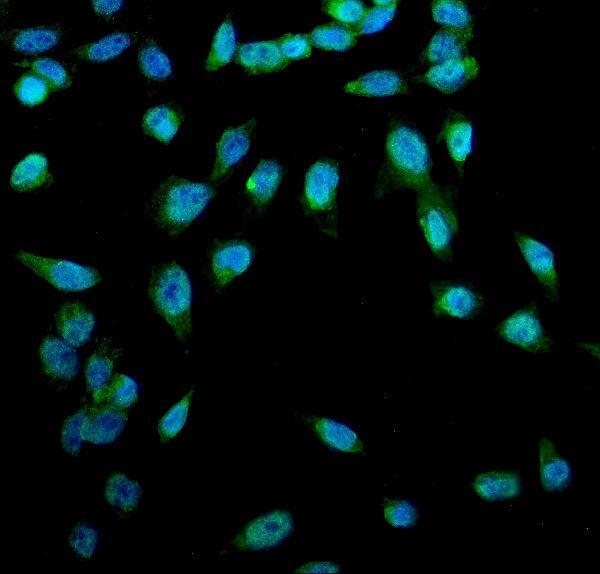
- Immunocytochemistry analysis of ACSL1 using anti-ACSL1antibody (Product # PA5-78713) . ACSL1 was detected in a section of U2OS cells. Enzyme antigen retrieval was performed using IHC enzyme antigen retrieval reagent for 15 mins. The cells were blocked with 10% goat serum and then incubated with 5μg/mL rabbit anti-ACSL1 antibody (Product # PA5-78713) overnight at 4°C. DyLight®488 Conjugated Goat Anti-Rabbit IgG was used as secondary antibody at 1:100 dilution and incubated for 30 minutes at 37°C. The section was counterstained with DAPI. Visualize using a fluorescence microscope and filter sets appropriate for the label used.
- Submitted by
- Invitrogen Antibodies (provider)
- Main image
- Experimental details
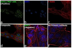
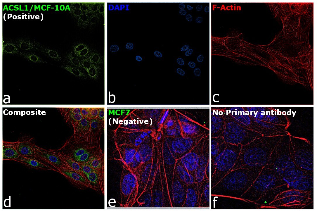
- Immunofluorescence analysis of ACSL1 was performed using 70% confluent log phase MCF-10A and MCF7 cells. The cells were fixed with 4% Paraformaldehyde for 10 minutes, permeabilized with 0.1% Triton™ X-100 for 10 minutes, and blocked with 2% BSA for 10 minutes at room temperature. The cells were labeled with ACSL1 Polyclonal Antibody (Product # PA5-78713) at 1:100 dilution in 0.1% BSA, incubated at 4 degree celsius overnight and then labeled with Goat anti-Rabbit IgG (H+L) Highly Cross-Adsorbed Secondary Antibody, Alexa Fluor Plus 488 (Product # A32731, 1:2000 dilution) for 45 minutes at room temperature (Panel a: Green). Nuclei (Panel b: Blue) were stained with SlowFade® Gold Antifade Mountant with DAPI (Product # S36938). F-actin (Panel c: Red) was stained with Rhodamine Phalloidin (Product # R415, 1:300). Panel d represents the merged image showing mitochondria like staining of ACSL1 in MCF-10A and not in MCF7 (panel e) which is reported negative for the same. Panel f represents control cells with no primary antibody to assess background. The images were captured at 60X magnification.
Supportive validation
- Submitted by
- Invitrogen Antibodies (provider)
- Main image
- Experimental details
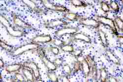
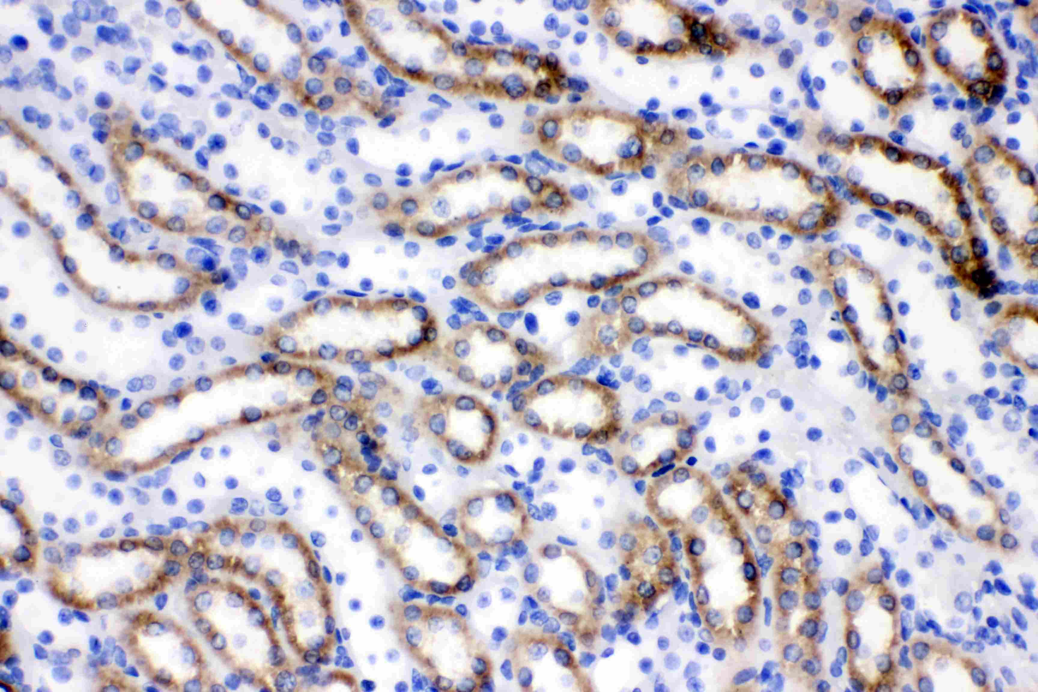
- Immunohistochemistry analysis of ACSL1 on paraffin-embedded rat kidney tissue. Sample was incubated with ACSL1 polyclonal antibody (Product# PA5-78713) with a dilution of 1 µg/mL, and developed by Streptavidin-Biotin-Complex (SABC) with DAB chromogen method.
- Submitted by
- Invitrogen Antibodies (provider)
- Main image

- Experimental details
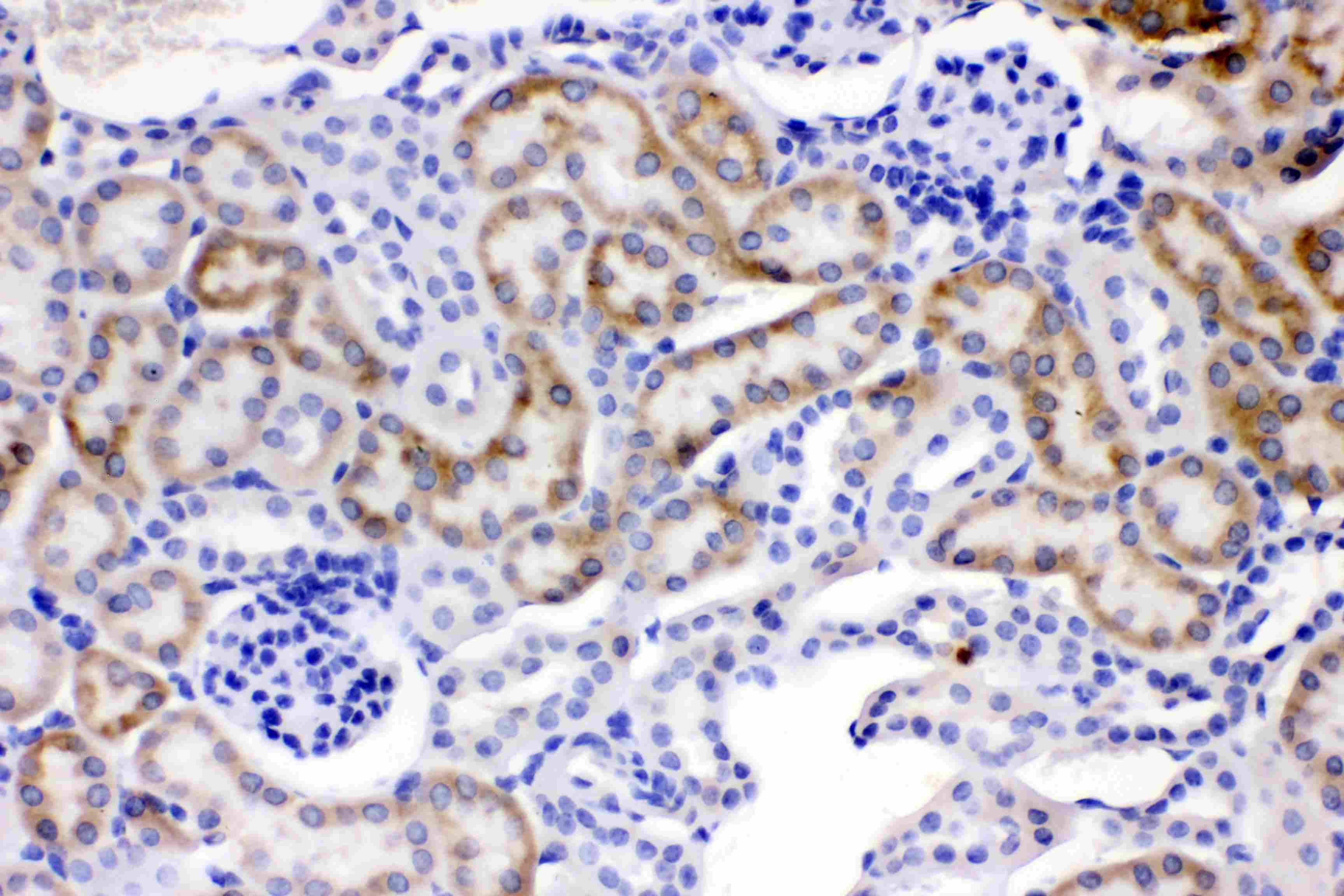
- Immunohistochemistry analysis of ACSL1 on paraffin-embedded mouse kidney tissue. Sample was incubated with ACSL1 polyclonal antibody (Product# PA5-78713) with a dilution of 1 µg/mL, and developed by Streptavidin-Biotin-Complex (SABC) with DAB chromogen method.
- Submitted by
- Invitrogen Antibodies (provider)
- Main image
- Experimental details
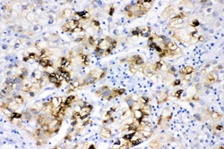
- Immunohistochemistry analysis of ACSL1 on paraffin-embedded human liver cancer tissue. Sample was incubated with ACSL1 polyclonal antibody (Product# PA5-78713) with a dilution of 1 µg/mL, and developed by Streptavidin-Biotin-Complex (SABC) with DAB chromogen method.

Supportive validation
- Submitted by
- Invitrogen Antibodies (provider)
- Main image
- Experimental details
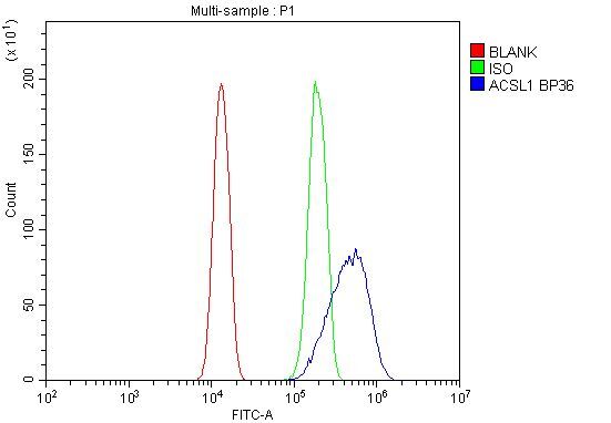
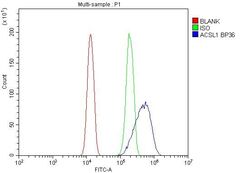
- Flow Cytometry of ACSL1 in A431 cells (blue line), isotype control rabbit IgG (green line) and unlabeled (red line). Samples were blocked with 10% goat serum, incubated with ACSL1 Polyclonal Antibody (Product # PA5-78713) at a dilution of 1 μg (per 1x10^6 cells), followed by DyLight®488 conjugated goat anti-rabbit IgG (for 30 minutes at 20°C) using 5-10 μg (per 1x10^6 cells) dilution.