Explore
 Validate
 Learn
Learn Western blot
Antibody data
- Antibody Data
- Antigen structure
- References [2]
- Comments [0]
- Validations
- Western blot [3]
- Immunocytochemistry [1]
- Immunohistochemistry [2]
Submit
Validation data
Reference
Comment
Report error
- Product number
- MA5-11626 - Provider product page

- Provider
- Invitrogen Antibodies
- Product name
- Caldesmon HMW Monoclonal Antibody (h-CALD)
- Antibody type
- Monoclonal
- Antigen
- Other
- Description
- MA5-11626 targets Caldesmon HMW in IHC (P), IF and WB applications and shows reactivity with human and mouse samples. The MA5-11626 immunogen is crude human uterus extract.
- Reactivity
- Human, Mouse, Rat
- Host
- Mouse
- Isotype
- IgG
- Antibody clone number
- h-CALD
- Vial size
- 500 µL
- Concentration
- 0.2 mg/mL
- Storage
- 4° C
Submitted references TGF-beta1 drives partial myofibroblastic differentiation in chondromyxoid fibroma of bone.
ALK-ATIC fusion in urinary bladder inflammatory myofibroblastic tumor.
Romeo S, Eyden B, Prins FA, Briaire-de Bruijn IH, Taminiau AH, Hogendoorn PC
The Journal of pathology 2006 Jan;208(1):26-34
The Journal of pathology 2006 Jan;208(1):26-34
ALK-ATIC fusion in urinary bladder inflammatory myofibroblastic tumor.
Debiec-Rychter M, Marynen P, Hagemeijer A, Pauwels P
Genes, chromosomes & cancer 2003 Oct;38(2):187-90
Genes, chromosomes & cancer 2003 Oct;38(2):187-90
No comments: Submit comment
Supportive validation
- Submitted by
- Invitrogen Antibodies (provider)
- Main image

- Experimental details
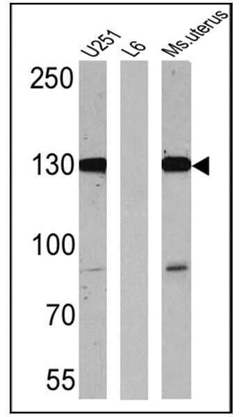
- Western blot analysis of Caldesmon HMW was performed by loading 25 µg of U251 (lane 1), L6 (lane 2) and mouse uterus (lane 3) onto an SDS polyacrylamide gel. Proteins were transferred to a PVDF membrane and blocked at 4ºC overnight. The membrane was probed with a Caldesmon HMW monoclonal antibody (Product # MA5-11626) at a dilution of 1:500 (U251 and mouse uterus) and 1:200 (L6) overnight at 4°C, washed in TBST, and probed with an HRP-conjugated secondary antibody for 1 hr at room temperature in the dark. Chemiluminescent detection was performed using Pierce ECL Plus Western Blotting Substrate (Product # 32132). Results show a band at ~130 kDa in mouse uterus and U251 lysates.
- Submitted by
- Invitrogen Antibodies (provider)
- Main image
- Experimental details
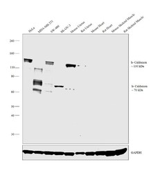
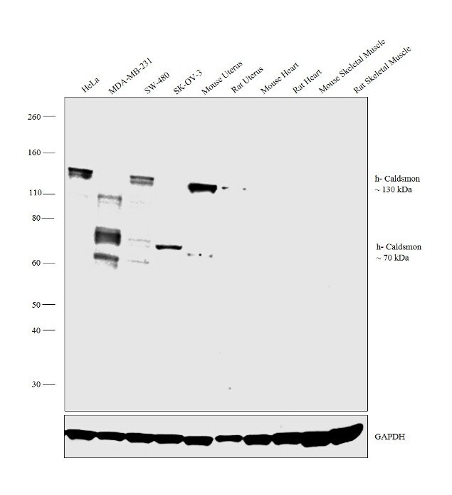
- Western blot analysis was performed on whole cell extracts (30 µg lysate) of HeLa (Lane 1), MDA-MB-231 (Lane 2), SW-480 (Lane 3), SK-OV-3 (Lane 4), tissue extracts of Mouse Uterus (Lane 5), Rat Uterus (Lane 6), Mouse Heart (Lane 7), Rat Heart (Lane 8), Mouse Skeletal Muscle (Lane 9) and Rat Skeletal Muscle (Lane 10). The blot was probed with Anti-Caldesmon HMW Monoclonal Antibody (Product # MA5-11626, 1:1000 dilution) and detected by chemiluminescence using Goat anti-Mouse IgG (H+L) Superclonal™ Secondary Antibody, HRP conjugate (Product # A28177, 0.25 µg/mL, 1:4000 dilution). A 130kDa band corresponding to h-Caldesmon (Caldesmon HMW) was observed in HeLa, SW-480, Mouse Uterus and Rat Uterus. A 70kDa band corresponding to l-Caldesmon (Caldesmon LMW) was observed in MDA-MB-231 and SK-OV-3. Both these bands were not presented in Mouse Heart, Rat Heart, Mouse Skeletal Muscle and Rat Skeletal Muscle which are reported negative for Caldesmon expression.
- Submitted by
- Invitrogen Antibodies (provider)
- Main image

- Experimental details
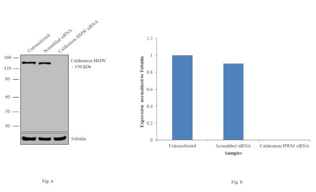
- Knockdown of Caldesmon HMW was achieved by transfecting HeLa cells with Caldesmon HMW specific siRNAs (Silencer® select Product # s2337, s2338). Western blot analysis (Fig. a) was performed using membrane extracts from the Caldesmon HMW knockdown cells (lane 3), non-specific scrambled siRNA transfected cells (lane 2) and untransfected cells (lane 1). The blots were probed with Caldesmon HMW Monoclonal Antibody (h-CALD) (Product # MA5-11626, 1:1000 dilution) and Goat anti-Mouse IgG (H+L) Superclonal™ Secondary Antibody, HRP conjugate (Product # A28177, 0.25 µg/mL, 1:4000 dilution). Densitometric analysis of this western blot is shown in histogram (Fig. b). Decrease in signal upon siRNA mediated knock down confirms that antibody is specific to Caldesmon HMW.
Supportive validation
- Submitted by
- Invitrogen Antibodies (provider)
- Main image
- Experimental details
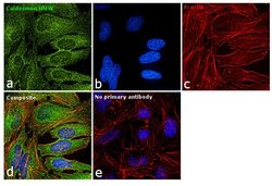
- Immunofluorescence analysis of Caldesmon was performed using 70% confluent log phase HeLa cells. The cells were fixed with 4% paraformaldehyde for 10 minutes, permeabilized with 0.1% Triton™ X-100 for 15 minutes, and blocked with 1% BSA for 1 hour at room temperature. The cells were labeled with Caldesmon HMW Monoclonal Antibody (h-CALD) (Product # MA5-11626) at 1:100 dilution in 0.1% BSA, incubated at 4 degree Celsius overnight and then labeled with Goat anti-Mouse IgG (H+L) Superclonal™ Secondary Antibody, Alexa Fluor® 488 conjugate (Product # A28175) at a dilution of 1:2000 for 45 minutes at room temperature (Panel a: green). Nuclei (Panel b: blue) were stained with ProLong™ Diamond Antifade Mountant with DAPI (Product # P36962). F-actin (Panel c: red) was stained with Rhodamine Phalloidin (Product # R415, 1:300). Panel d represents the merged image showing cytoskeletal localization. Panel e represents control cells with no primary antibody to assess background. The images were captured at 60X magnification.

Supportive validation
- Submitted by
- Invitrogen Antibodies (provider)
- Main image
- Experimental details
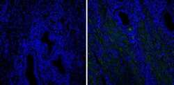
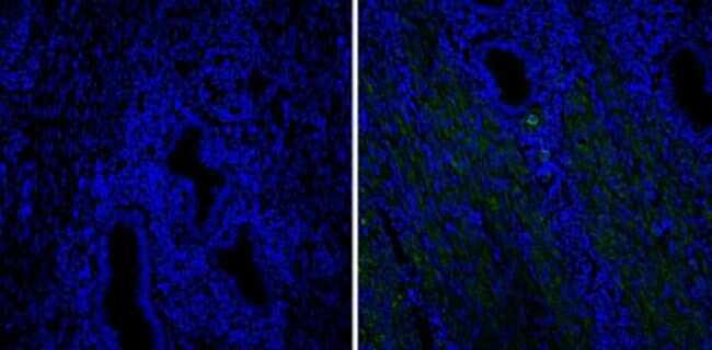
- Immunofluorescent analysis of Caldesmon HMW (green) showing staining in human uterus tissue (right) compared to a negative control without primary antibody (left). Formalin-fixed cells were permeabilized with 0.1% Triton X-100 in TBS for 5-10 minutes and blocked with 3% BSA-PBS for 30 minutes at room temperature. Cells were probed with a Caldesmon HMW monoclonal antibody (Product # MA5-11626) in 3% BSA-PBS at a dilution of 1:20 and incubated overnight at 4ºC in a humidified chamber. Cells were washed with PBST and incubated with a DyLight-488 conjugated secondary antibody in PBS at room temperature in the dark. Nuclei were stained with DAPI (blue) and images were taken at a magnification of 60x.
- Submitted by
- Invitrogen Antibodies (provider)
- Main image
- Experimental details
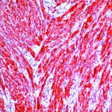
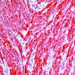
- Formalin-fixed, paraffin-embedded human uterus stained with Caldesmon antibody using peroxidase-conjugate and AEC chromogen. Note cytoplasmic staining of smooth muscles.