Explore
 Validate
 Learn
Learn Western blot
 Immunocytochemistry
Antibody data
- Antibody Data
- Antigen structure
- References [4]
- Comments [0]
- Validations
- Western blot [4]
- Immunohistochemistry [8]
Submit
Validation data
Reference
Comment
Report error
- Product number
- NBP1-85702 - Provider product page

- Provider
- Novus Biologicals
- Proper citation
- Novus Cat#NBP1-85702, RRID:AB_11039260
- Product name
- Rabbit Polyclonal Caldesmon/CALD1 Antibody
- Antibody type
- Polyclonal
- Description
- Immunogen affinity purified. Specificity of human Caldesmon/CALD1 antibody verified on a Protein Array containing target protein plus 383 other non-specific proteins.
- Reactivity
- Human, Mouse, Rat
- Host
- Rabbit
- Isotype
- IgG
- Vial size
- 0.1 ml
- Storage
- Store at 4C short term. Aliquot and store at -20C long term. Avoid freeze-thaw cycles.
Submitted references Stromal gene expression defines poor-prognosis subtypes in colorectal cancer.
Systematic validation of antibody binding and protein subcellular localization using siRNA and confocal microscopy.
Histochemical localization of caldesmon in the CNS and ganglia of the mouse.
Mapping the subcellular protein distribution in three human cell lines.
Calon A, Lonardo E, Berenguer-Llergo A, Espinet E, Hernando-Momblona X, Iglesias M, Sevillano M, Palomo-Ponce S, Tauriello DV, Byrom D, Cortina C, Morral C, Barceló C, Tosi S, Riera A, Attolini CS, Rossell D, Sancho E, Batlle E
Nature genetics 2015 Apr;47(4):320-9
Nature genetics 2015 Apr;47(4):320-9
Systematic validation of antibody binding and protein subcellular localization using siRNA and confocal microscopy.
Stadler C, Hjelmare M, Neumann B, Jonasson K, Pepperkok R, Uhlén M, Lundberg E
Journal of proteomics 2012 Apr 3;75(7):2236-51
Journal of proteomics 2012 Apr 3;75(7):2236-51
Histochemical localization of caldesmon in the CNS and ganglia of the mouse.
Köhler CN
The journal of histochemistry and cytochemistry : official journal of the Histochemistry Society 2011 May;59(5):504-17
The journal of histochemistry and cytochemistry : official journal of the Histochemistry Society 2011 May;59(5):504-17
Mapping the subcellular protein distribution in three human cell lines.
Fagerberg L, Stadler C, Skogs M, Hjelmare M, Jonasson K, Wiking M, Abergh A, Uhlén M, Lundberg E
Journal of proteome research 2011 Aug 5;10(8):3766-77
Journal of proteome research 2011 Aug 5;10(8):3766-77
No comments: Submit comment
Supportive validation
- Submitted by
- Novus Biologicals (provider)
- Main image
- Experimental details
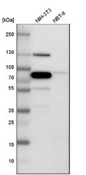
- Western Blot: Caldesmon/CALD1 Antibody [NBP1-85702] - Analysis in mouse cell line NIH-3T3 and rat cell line NBT-II.

- Submitted by
- Novus Biologicals (provider)
- Main image
- Experimental details
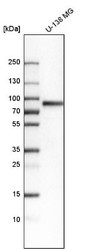
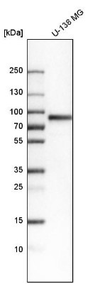
- Western Blot: Caldesmon/CALD1 Antibody [NBP1-85702] - Analysis in human cell line U-138 MG.
- Submitted by
- Novus Biologicals (provider)
- Main image
- Experimental details
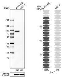
- Western Blot: Caldesmon/CALD1 Antibody [NBP1-85702] - Analysis in human cell lines U-251MG and MCF-7. Corresponding RNA-seq data are presented for the same cell lines. Loading control: Anti-GAPDH.

- Submitted by
- Novus Biologicals (provider)
- Main image
- Experimental details
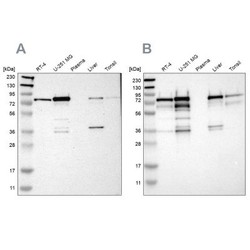
- Western Blot: Caldesmon/CALD1 Antibody [NBP1-85702] - Analysis using Anti-CALD1 antibody NBP1-85702 (A) shows similar pattern to independent antibody NBP1-85701 (B).

Supportive validation
- Submitted by
- Novus Biologicals (provider)
- Main image
- Experimental details
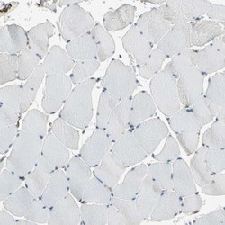
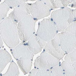
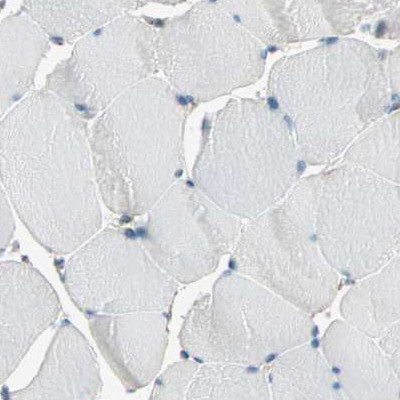
- Immunohistochemistry-Paraffin: Caldesmon/CALD1 Antibody [NBP1-85702] - Staining of human skeletal muscle shows low expression as expected.
- Submitted by
- Novus Biologicals (provider)
- Main image
- Experimental details
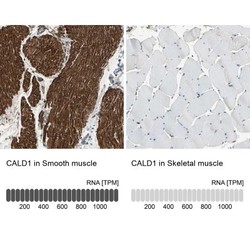
- Immunohistochemistry-Paraffin: Caldesmon/CALD1 Antibody [NBP1-85702] - Staining in human smooth muscle and skeletal muscle tissues using anti-CALD1 antibody. Corresponding CALD1 RNA-seq data are presented for the same tissues.

- Submitted by
- Novus Biologicals (provider)
- Main image
- Experimental details
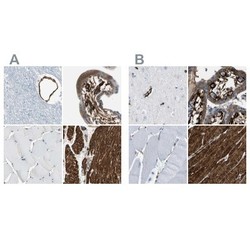
- Immunohistochemistry-Paraffin: Caldesmon/CALD1 Antibody [NBP1-85702] - Staining of human cerebral cortex, placenta, skeletal muscle and smooth muscle using Anti-CALD1 antibody NBP1-85702 (A) shows similar protein distribution across tissues to independent antibody NBP1-85701 (B).

- Submitted by
- Novus Biologicals (provider)
- Main image
- Experimental details
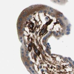
- Immunohistochemistry-Paraffin: Caldesmon/CALD1 Antibody [NBP1-85702] - Staining of human placenta.

- Submitted by
- Novus Biologicals (provider)
- Main image
- Experimental details
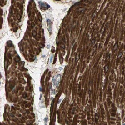
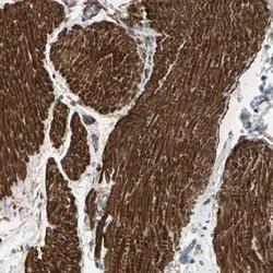
- Immunohistochemistry: Caldesmon/CALD1 Antibody [NBP1-85702] - Immunohistochemical staining of human smooth muscle shows high expression.

- Submitted by
- Novus Biologicals (provider)
- Main image

- Experimental details
- Immunohistochemistry-Paraffin: Caldesmon/CALD1 Antibody [NBP1-85702] - Staining of human cerebral cortex.
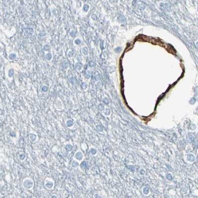
- Submitted by
- Novus Biologicals (provider)
- Main image
- Experimental details
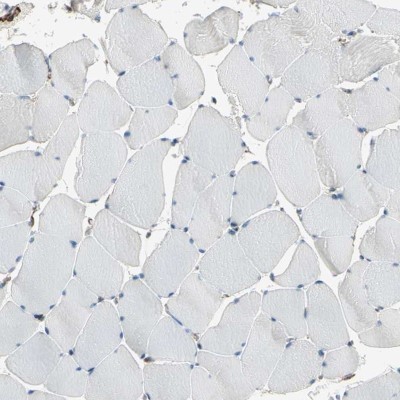
- Immunohistochemistry-Paraffin: Caldesmon/CALD1 Antibody [NBP1-85702] - Staining of human skeletal muscle using Anti-CALD1 antibody NBP1-85702.
- Submitted by
- Novus Biologicals (provider)
- Main image

- Experimental details
- Immunohistochemistry-Paraffin: Caldesmon/CALD1 Antibody [NBP1-85702] - Staining of human smooth muscle using Anti-CALD1 antibody NBP1-85702.