 Explore
 Validate
 Learn
Learn Western blot
 Immunocytochemistry
Antibody data
- Antibody Data
- Antigen structure
- References [7]
- Comments [0]
- Validations
- Immunocytochemistry [1]
- Immunohistochemistry [1]
Submit
Validation data
Reference
Comment
Report error
- Product number
- HPA008066 - Provider product page

- Provider
- Atlas Antibodies
- Proper citation
- Atlas Antibodies Cat#HPA008066, RRID:AB_1078378
- Product name
- Anti-CALD1
- Antibody type
- Polyclonal
- Description
- Polyclonal Antibody against Human CALD1, Gene description: caldesmon 1, Alternative Gene Names: CDM, H-CAD, L-CAD, Validated applications: ICC, WB, IHC, Uniprot ID: Q05682, Storage: Store at +4°C for short term storage. Long time storage is recommended at -20°C.
- Reactivity
- Human, Mouse, Rat
- Host
- Rabbit
- Conjugate
- Unconjugated
- Isotype
- IgG
- Vial size
- 100 µl
- Concentration
- 0.1 mg/ml
- Storage
- Store at +4°C for short term storage. Long time storage is recommended at -20°C.
- Handling
- The antibody solution should be gently mixed before use.
Submitted references Epithelial NOTCH Signaling Rewires the Tumor Microenvironment of Colorectal Cancer to Drive Poor-Prognosis Subtypes and Metastasis
TGFβ drives immune evasion in genetically reconstituted colon cancer metastasis
Stromal gene expression defines poor-prognosis subtypes in colorectal cancer
Stromal gene expression defines poor-prognosis subtypes in colorectal cancer
Systematic validation of antibody binding and protein subcellular localization using siRNA and confocal microscopy
Mapping the Subcellular Protein Distribution in Three Human Cell Lines
Histochemical localization of caldesmon in the CNS and ganglia of the mouse.
Jackstadt R, van Hooff S, Leach J, Cortes-Lavaud X, Lohuis J, Ridgway R, Wouters V, Roper J, Kendall T, Roxburgh C, Horgan P, Nixon C, Nourse C, Gunzer M, Clark W, Hedley A, Yilmaz O, Rashid M, Bailey P, Biankin A, Campbell A, Adams D, Barry S, Steele C, Medema J, Sansom O
Cancer Cell 2019;36(3):319-336.e7
Cancer Cell 2019;36(3):319-336.e7
TGFβ drives immune evasion in genetically reconstituted colon cancer metastasis
Tauriello D, Palomo-Ponce S, Stork D, Berenguer-Llergo A, Badia-Ramentol J, Iglesias M, Sevillano M, Ibiza S, Cañellas A, Hernando-Momblona X, Byrom D, Matarin J, Calon A, Rivas E, Nebreda A, Riera A, Attolini C, Batlle E
Nature 2018;554(7693):538-543
Nature 2018;554(7693):538-543
Stromal gene expression defines poor-prognosis subtypes in colorectal cancer
Calon A, Lonardo E, Berenguer-Llergo A, Espinet E, Hernando-Momblona X, Iglesias M, Sevillano M, Palomo-Ponce S, Tauriello D, Byrom D, Cortina C, Morral C, Barceló C, Tosi S, Riera A, Attolini C, Rossell D, Sancho E, Batlle E
Nature Genetics 2015;47(4):320-329
Nature Genetics 2015;47(4):320-329
Stromal gene expression defines poor-prognosis subtypes in colorectal cancer
Calon A, Lonardo E, Berenguer-Llergo A, Espinet E, Hernando-Momblona X, Iglesias M, Sevillano M, Palomo-Ponce S, Tauriello D, Byrom D, Cortina C, Morral C, Barceló C, Tosi S, Riera A, Attolini C, Rossell D, Sancho E, Batlle E
Nature Genetics 2015 February;47(4):320-329
Nature Genetics 2015 February;47(4):320-329
Systematic validation of antibody binding and protein subcellular localization using siRNA and confocal microscopy
Stadler C, Hjelmare M, Neumann B, Jonasson K, Pepperkok R, Uhlén M, Lundberg E
Journal of Proteomics 2012;75(7):2236-2251
Journal of Proteomics 2012;75(7):2236-2251
Mapping the Subcellular Protein Distribution in Three Human Cell Lines
Fagerberg L, Stadler C, Skogs M, Hjelmare M, Jonasson K, Wiking M, Åbergh A, Uhlén M, Lundberg E
Journal of Proteome Research 2011;10(8):3766-3777
Journal of Proteome Research 2011;10(8):3766-3777
Histochemical localization of caldesmon in the CNS and ganglia of the mouse.
Köhler CN
The journal of histochemistry and cytochemistry : official journal of the Histochemistry Society 2011 May;59(5):504-17
The journal of histochemistry and cytochemistry : official journal of the Histochemistry Society 2011 May;59(5):504-17
No comments: Submit comment
Supportive validation
- Submitted by
- Atlas Antibodies (provider)
- Main image
- Experimental details
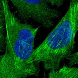
- Immunofluorescent staining of human cell line U-2 OS shows localization to plasma membrane & actin filaments.
- Sample type
- Human

Supportive validation
- Submitted by
- Atlas Antibodies (provider)
- Enhanced method
- Orthogonal validation
- Main image
- Experimental details
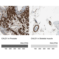
- Immunohistochemistry analysis in human prostate and skeletal muscle tissues using HPA008066 antibody. Corresponding CALD1 RNA-seq data are presented for the same tissues.
- Sample type
- Human
- Protocol
- Protocol
