 Explore
 Validate
 Learn
Learn Western blot
 Immunohistochemistry
Antibody data
- Antibody Data
- Antigen structure
- References [4]
- Comments [0]
- Validations
- Immunohistochemistry [1]
Submit
Validation data
Reference
Comment
Report error
- Product number
- HPA007493 - Provider product page

- Provider
- Atlas Antibodies
- Proper citation
- Atlas Antibodies Cat#HPA007493, RRID:AB_1848642
- Product name
- Anti-WIPI1
- Antibody type
- Polyclonal
- Description
- Polyclonal Antibody against Human WIPI1, Gene description: WD repeat domain, phosphoinositide interacting 1, Alternative Gene Names: Atg18, ATG18A, FLJ10055, WIPI49, Validated applications: WB, IHC, Uniprot ID: Q5MNZ9, Storage: Store at +4°C for short term storage. Long time storage is recommended at -20°C.
- Reactivity
- Human, Mouse, Rat
- Host
- Rabbit
- Conjugate
- Unconjugated
- Isotype
- IgG
- Vial size
- 100 µl
- Concentration
- 0.1 mg/ml
- Storage
- Store at +4°C for short term storage. Long time storage is recommended at -20°C.
- Handling
- The antibody solution should be gently mixed before use.
Submitted references Mediobasal hypothalamic FKBP51 acts as a molecular switch linking autophagy to whole-body metabolism
Enhanced autophagic-lysosomal activity and increased BAG3-mediated selective macroautophagy as adaptive response of neuronal cells to chronic oxidative stress
WIPI1, BAG1, and PEX3 Autophagy‐Related Genes Are Relevant Melanoma Markers
Estrogen receptor α regulates non-canonical autophagy that provides stress resistance to neuroblastoma and breast cancer cells and involves BAG3 function
Häusl A, Bajaj T, Brix L, Pöhlmann M, Hafner K, De Angelis M, Nagler J, Dethloff F, Balsevich G, Schramm K, Giavalisco P, Chen A, Schmidt M, Gassen N
Science Advances 2022;8(10)
Science Advances 2022;8(10)
Enhanced autophagic-lysosomal activity and increased BAG3-mediated selective macroautophagy as adaptive response of neuronal cells to chronic oxidative stress
Chakraborty D, Felzen V, Hiebel C, Stürner E, Perumal N, Manicam C, Sehn E, Grus F, Wolfrum U, Behl C
Redox Biology 2019;24
Redox Biology 2019;24
WIPI1, BAG1, and PEX3 Autophagy‐Related Genes Are Relevant Melanoma Markers
D’Arcangelo D, Giampietri C, Muscio M, Scatozza F, Facchiano F, Facchiano A, Sanchez-Alvarez M
Oxidative Medicine and Cellular Longevity 2018;2018(1)
Oxidative Medicine and Cellular Longevity 2018;2018(1)
Estrogen receptor α regulates non-canonical autophagy that provides stress resistance to neuroblastoma and breast cancer cells and involves BAG3 function
Felzen V, Hiebel C, Koziollek-Drechsler I, Reißig S, Wolfrum U, Kögel D, Brandts C, Behl C, Morawe T
Cell Death & Disease 2015;6(7):e1812-e1812
Cell Death & Disease 2015;6(7):e1812-e1812
No comments: Submit comment
Supportive validation
- Submitted by
- Atlas Antibodies (provider)
- Enhanced method
- Orthogonal validation
- Main image
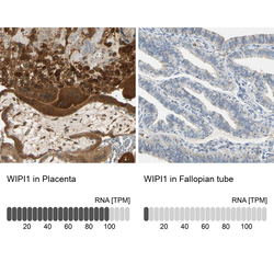
- Experimental details
- Immunohistochemistry analysis in human placenta and fallopian tube tissues using HPA007493 antibody. Corresponding WIPI1 RNA-seq data are presented for the same tissues.
- Sample type
- Human
- Protocol
- Protocol
