 Explore
 Validate
 Learn
Learn Western blot
 Immunocytochemistry
Antibody data
- Antibody Data
- Antigen structure
- References [1]
- Comments [0]
- Validations
- Immunocytochemistry [1]
- Immunohistochemistry [1]
- Other assay [1]
Submit
Validation data
Reference
Comment
Report error
- Product number
- PA5-28584 - Provider product page

- Provider
- Invitrogen Antibodies
- Product name
- SPHK1 Polyclonal Antibody
- Antibody type
- Polyclonal
- Antigen
- Recombinant full-length protein
- Description
- Recommended positive controls: 293T, A431, H1299, HeLaS3, HepG2, Molt-4, Raji. Predicted reactivity: Human (99%). Store product as a concentrated solution. Centrifuge briefly prior to opening the vial.
- Reactivity
- Human
- Host
- Rabbit
- Isotype
- IgG
- Vial size
- 100 μL
- Concentration
- 1 mg/mL
- Storage
- Store at 4°C short term. For long term storage, store at -20°C, avoiding freeze/thaw cycles.
Submitted references Mfsd2a and Spns2 are essential for sphingosine-1-phosphate transport in the formation and maintenance of the blood-brain barrier.
Wang Z, Zheng Y, Wang F, Zhong J, Zhao T, Xie Q, Zhu T, Ma F, Tang Q, Zhou B, Zhu J
Science advances 2020 May;6(22):eaay8627
Science advances 2020 May;6(22):eaay8627
No comments: Submit comment
Supportive validation
- Submitted by
- Invitrogen Antibodies (provider)
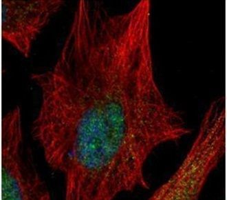
- Main image

- Experimental details
- Immunofluorescent analysis of SPHK1 in paraformaldehyde-fixed HeLa cells using a SPHK1 polyclonal antibody (Product # PA5-28584) (Green) at a 1:500 dilution. Alpha-tubulin filaments were labeled with Product # PA5-29281 (Red) at a 1:2000.
Supportive validation
- Submitted by
- Invitrogen Antibodies (provider)
- Main image
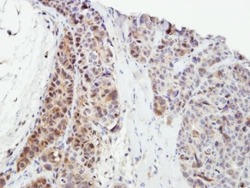
- Experimental details
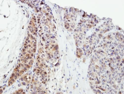
- Immunohistochemical analysis of paraffin-embedded SW480 xenograft, using SPHK1 (Product # PA5-28584) antibody at 1:500 dilution. Antigen Retrieval: EDTA based buffer, pH 8.0, 15 min.
Supportive validation
- Submitted by
- Invitrogen Antibodies (provider)
- Main image
- Experimental details
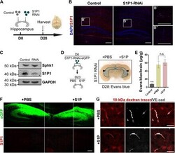
- Fig. 3 The S1P signaling pathway is crucial for the maintenance of the BBB. Knock down of S1P1 in the hippocampus led to BBB breakdown. ( A ) Strategy used for S1P1-RNAi transfection. ( B ) Immunostaining for S1P1 showed that the expression of S1P1 was inhibited after RNAi transfection. ( C ) Western blot showing that S1P1 knockdown had no substantial effect on the expression of Sphk1 at D28. ( D ) S1P or PBS was added in situ at 23 days post AAV injection; Evans blue leakage was confined to the hippocampus (black arrowheads). ( E ) Quantification of Evans blue staining showed that additional S1P had no effect on BBB breakdown. ( F ) The inhibition of S1P1 was not affected by additional PBS or S1P. ( G ) The tracer penetrated into the brain parenchyma, indicating that the BBB did not recover after adding S1P. Scale bars: 200 mum in B and F; 100 mum in G. Error bars: SEM. Significance determined by Students t -test: *** P < 0.001, n.s. P > 0.05. Each image is representative of three individual male mice. Photo credit: Fan Wang, State Key Laboratory of Medical Neurobiology, the Institutes of Brain Science and the Collaborative Innovation Center for Brain Science, Shanghai Medical College, Fudan University.
