Explore
 Validate
 Learn
Learn Western blot
 Immunoprecipitation
Antibody data
- Antibody Data
- Antigen structure
- References [1]
- Comments [0]
- Validations
- Western blot [1]
- ELISA [1]
- Immunocytochemistry [1]
- Immunohistochemistry [1]
Submit
Validation data
Reference
Comment
Report error
- Product number
- AF5536 - Provider product page

- Provider
- R&D Systems
- Product name
- Human Sphingosine Kinase 1/SPHK1 Antibody
- Antibody type
- Polyclonal
- Description
- Antigen Affinity-purified. Detects human SPHK1 in direct ELISAs and Western blots. In direct ELISAs, less than 1% cross-reactivity with recombinant human SPHK2 is observed.
- Reactivity
- Human
- Host
- Sheep
- Conjugate
- Unconjugated
- Antigen sequence
NP_068807- Isotype
- IgG
- Vial size
- 100 ug
- Concentration
- LYOPH
- Storage
- Use a manual defrost freezer and avoid repeated freeze-thaw cycles. 12 months from date of receipt, -20 to -70 °C as supplied. 1 month, 2 to 8 °C under sterile conditions after reconstitution. 6 months, -20 to -70 °C under sterile conditions after reconstitution.
Submitted references Pseudomonas-derived ceramidase induces production of inflammatory mediators from human keratinocytes via sphingosine-1-phosphate.
Oizumi A, Nakayama H, Okino N, Iwahara C, Kina K, Matsumoto R, Ogawa H, Takamori K, Ito M, Suga Y, Iwabuchi K
PloS one 2014;9(2):e89402
PloS one 2014;9(2):e89402
No comments: Submit comment
Supportive validation
- Submitted by
- R&D Systems (provider)
- Main image
- Experimental details
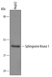
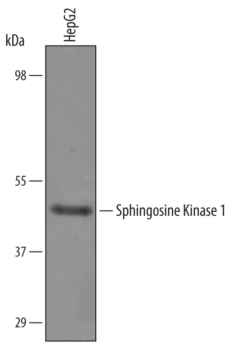
- Detection of Human Sphingosine Kinase 1/SPHK1 by Western Blot. Western blot shows lysates of HepG2 human hepatocellular carcinoma cell line. PVDF Membrane was probed with 0.5 µg/mL of Sheep Anti-Human Sphingosine Kinase 1/SPHK1 Antigen Affinity-purified Polyclonal Antibody (Catalog # AF5536) followed by HRP-conjugated Anti-Sheep IgG Secondary Antibody (Catalog # HAF016). A specific band was detected for Sphingosine Kinase 1/SPHK1 at approximately 45 kDa (as indicated). This experiment was conducted under reducing conditions and using Immunoblot Buffer Group 8.
Supportive validation
- Submitted by
- R&D Systems (provider)
- Main image
- Experimental details
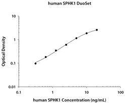
- Human Sphingosine Kinase 1/SPHK1 ELISA Standard Curve. Recombinant Human Sphingosine Kinase 1/SPHK1 protein was serially diluted 2-fold and captured by Mouse Anti-Human Sphingosine Kinase 1/SPHK1 Monoclonal Antibody (Catalog # MAB5536) coated on a Clear Polystyrene Microplate (Catalog # DY990). Sheep Anti-Human Sphingosine Kinase 1/SPHK1 Antigen Affinity-purified Polyclonal Antibody (Catalog # AF5536) was biotinylated and incubated with the protein captured on the plate. Detection of the standard curve was achieved by incubating Streptavidin-HRP (Catalog # DY998) followed by Substrate Solution (Catalog # DY999) and stopping the enzymatic reaction with Stop Solution (Catalog # DY994).

Supportive validation
- Submitted by
- R&D Systems (provider)
- Main image

- Experimental details
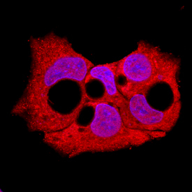
- Sphingosine Kinase 1/SPHK1 in HepG2 Human Cell Line. Sphingosine Kinase 1/SPHK1 was detected in immersion fixed HepG2 human hepatocellular carcinoma cell line using Sheep Anti-Human Sphingosine Kinase 1/SPHK1 Antigen Affinity-purified Polyclonal Antibody (Catalog # AF5536) at 10 µg/mL for 3 hours at room temperature. Cells were stained using the NorthernLights™ 557-conjugated Anti-Sheep IgG Secondary Antibody (red; Catalog # NL010) and counterstained with DAPI (blue). Specific staining was localized to cytoplasm. View our protocol for Fluorescent ICC Staining of Cells on Coverslips.
Supportive validation
- Submitted by
- R&D Systems (provider)
- Main image

- Experimental details
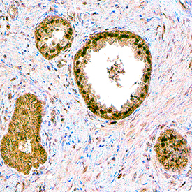
- Sphingosine Kinase 1/SPHK1 in Human Prostate. Sphingosine Kinase 1/SPHK1 was detected in immersion fixed paraffin-embedded sections of human prostate using Sheep Anti-Human Sphingosine Kinase 1/SPHK1 Antigen Affinity-purified Polyclonal Antibody (Catalog # AF5536) at 15 µg/mL overnight at 4 °C. Tissue was stained using the Anti-Sheep HRP-DAB Cell & Tissue Staining Kit (brown; Catalog # CTS019) and counterstained with hematoxylin (blue). Specific staining was localized to cytoplasm and nuclei of epithelial cells. View our protocol for Chromogenic IHC Staining of Paraffin-embedded Tissue Sections.