Explore
 Validate
 Learn
Learn Western blot
 ELISA
ELISAAntibody data
- Antibody Data
- Antigen structure
- References [8]
- Comments [0]
- Validations
- Western blot [1]
- Immunohistochemistry [1]
Submit
Validation data
Reference
Comment
Report error
- Product number
- 12442-2-AP - Provider product page

- Provider
- Proteintech Group
- Proper citation
- Proteintech Cat#12442-2-AP, RRID:AB_2162893
- Product name
- PDE1A antibody
- Antibody type
- Polyclonal
- Description
- KD/KO validated PDE1A antibody (Cat. #12442-2-AP) is a rabbit polyclonal antibody that shows reactivity with human, mouse and has been validated for the following applications: IHC, IP, WB, ELISA.
- Reactivity
- Human, Mouse
- Host
- Rabbit
- Conjugate
- Unconjugated
- Isotype
- IgG
- Vial size
- 20ul, 150ul
Submitted references Phosphodiesterase 1A overexpression contributes to the progression of renal fibrosis.
Therapy-induced senescence is a transient drug resistance mechanism in breast cancer.
Phosphodiesterase 1A physically interacts with YTHDF2 and reinforces the progression of non-small cell lung cancer.
Transcriptome profiling of intact bowel wall reveals that PDE1A and SEMA3D are possible markers with roles in enteric smooth muscle apoptosis, proliferative disorders, and dysautonomia in Crohn's disease.
Single-Cell Analysis of Foxp1-Driven Mechanisms Essential for Striatal Development.
Follicle-stimulating hormone and luteinizing hormone increase Ca2+ in the granulosa cells of mouse ovarian follicles†.
Generation and phenotypic characterization of Pde1a mutant mice.
Luteinizing Hormone Causes Phosphorylation and Activation of the cGMP Phosphodiesterase PDE5 in Rat Ovarian Follicles, Contributing, Together with PDE1 Activity, to the Resumption of Meiosis.
Hong W, Zhao B, Wu W, Song A, Wang M, Lu J, Li F, Lu R, Dai H, Xie K, Min L, Gu L
Molecular therapy : the journal of the American Society of Gene Therapy 2026 Mar 4;34(3):1539-1553
Molecular therapy : the journal of the American Society of Gene Therapy 2026 Mar 4;34(3):1539-1553
Therapy-induced senescence is a transient drug resistance mechanism in breast cancer.
Bajtai E, Kiss C, Bakos É, Langó T, Lovrics A, Schád É, Tisza V, Hegedűs K, Fürjes P, Szabó Z, Tusnády GE, Szakács G, Tantos Á, Spisák S, Tóvári J, Füredi A
Molecular cancer 2025 May 1;24(1):128
Molecular cancer 2025 May 1;24(1):128
Phosphodiesterase 1A physically interacts with YTHDF2 and reinforces the progression of non-small cell lung cancer.
Zhang C, Zhang Z, Wu Y, Wu Y, Cheng J, Luo K, Li Z, Zhang M, Wang J, Zhang X, Li Y
eLife 2025 Jul 24;13
eLife 2025 Jul 24;13
Transcriptome profiling of intact bowel wall reveals that PDE1A and SEMA3D are possible markers with roles in enteric smooth muscle apoptosis, proliferative disorders, and dysautonomia in Crohn's disease.
Yang Y, Xia L, Yang W, Wang Z, Meng W, Zhang M, Ma Q, Gou J, Wang J, Shu Y, Wu X
Frontiers in genetics 2023;14:1194882
Frontiers in genetics 2023;14:1194882
Single-Cell Analysis of Foxp1-Driven Mechanisms Essential for Striatal Development.
Anderson AG, Kulkarni A, Harper M, Konopka G
Cell reports 2020 Mar 3;30(9):3051-3066.e7
Cell reports 2020 Mar 3;30(9):3051-3066.e7
Follicle-stimulating hormone and luteinizing hormone increase Ca2+ in the granulosa cells of mouse ovarian follicles†.
Egbert JR, Fahey PG, Reimer J, Owen CM, Evsikov AV, Nikolaev VO, Griesbeck O, Ray RS, Tolias AS, Jaffe LA
Biology of reproduction 2019 Aug 1;101(2):433-444
Biology of reproduction 2019 Aug 1;101(2):433-444
Generation and phenotypic characterization of Pde1a mutant mice.
Wang X, Yamada S, LaRiviere WB, Ye H, Bakeberg JL, Irazabal MV, Chebib FT, van Deursen J, Harris PC, Sussman CR, Behfar A, Ward CJ, Torres VE
PloS one 2017;12(7):e0181087
PloS one 2017;12(7):e0181087
Luteinizing Hormone Causes Phosphorylation and Activation of the cGMP Phosphodiesterase PDE5 in Rat Ovarian Follicles, Contributing, Together with PDE1 Activity, to the Resumption of Meiosis.
Egbert JR, Uliasz TF, Shuhaibar LC, Geerts A, Wunder F, Kleiman RJ, Humphrey JM, Lampe PD, Artemyev NO, Rybalkin SD, Beavo JA, Movsesian MA, Jaffe LA
Biology of reproduction 2016 May;94(5):110
Biology of reproduction 2016 May;94(5):110
No comments: Submit comment
Supportive validation
- Submitted by
- Proteintech Group (provider)
- Main image
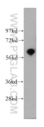
- Experimental details
- human brain tissue were subjected to SDS PAGE followed by western blot with 12442-2-AP(PDE1A antibody) at dilution of 1:500
- Sample type
- tissue

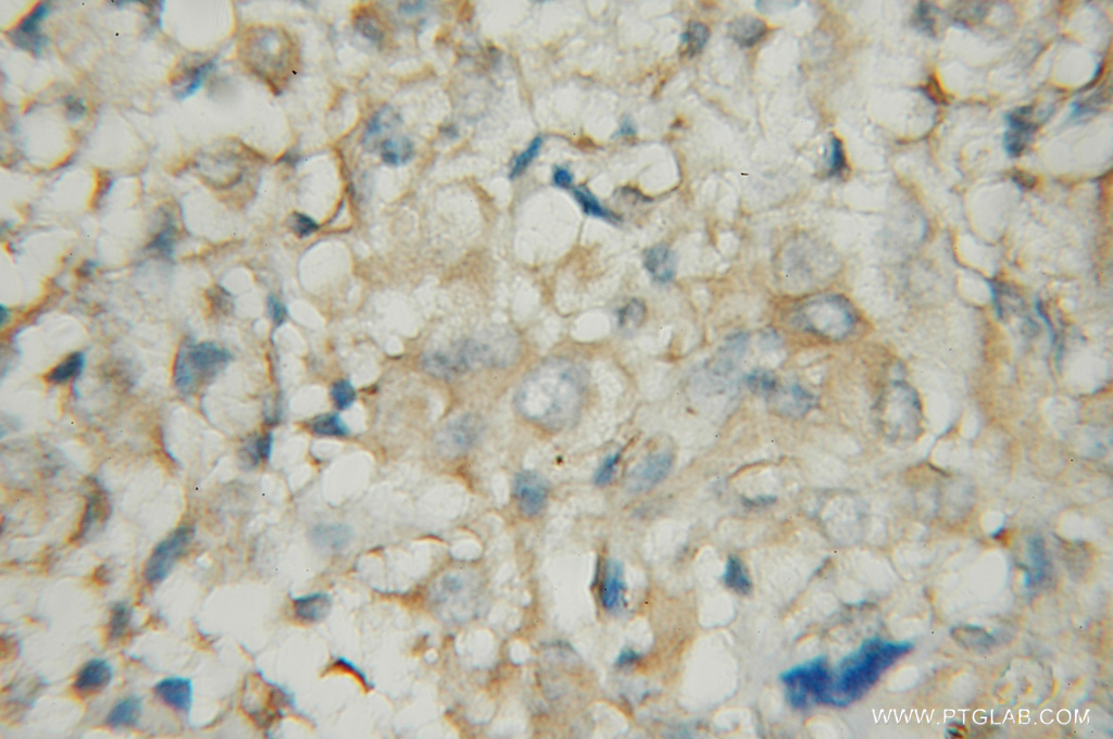
Supportive validation
- Submitted by
- Proteintech Group (provider)
- Main image
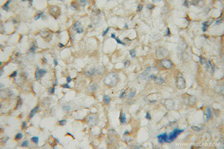
- Experimental details
- The PDE1A antibody from Proteintech is a rabbit polyclonal antibody to a recombinant protein of human PDE1A. This antibody recognizes human, mouse antigen. The PDE1A antibody has been validated for the following applications: ELISA, WB, IHC analysis.