 Explore
 Validate
 Learn
Learn Western blot
 ELISA
ELISAAntibody data
- Antibody Data
- Antigen structure
- References [5]
- Comments [0]
- Validations
- Western blot [4]
- Immunohistochemistry [2]
Submit
Validation data
Reference
Comment
Report error
- Product number
- GTX20292 - Provider product page

- Provider
- GeneTex
- Proper citation
- GeneTex Cat#GTX20292, RRID:AB_384293
- Product name
- Collagen I antibody
- Antibody type
- Polyclonal
- Reactivity
- Human, Mouse, Rat, Bovine
- Host
- Rabbit
Submitted references Bilateral sympathetic stellate ganglionectomy attenuates myocardial remodelling and fibrosis in a rat model of chronic volume overload.
Autologous Adipose-Derived Tissue Matrix Part I: Biologic Characteristics.
The Role of Butylidenephthalide in Targeting the Microenvironment Which Contributes to Liver Fibrosis Amelioration.
Saikosaponin d induces cell death through caspase-3-dependent, caspase-3-independent and mitochondrial pathways in mammalian hepatic stellate cells.
Lyso-globotriaosylceramide downregulates KCa3.1 channel expression to inhibit collagen synthesis in fibroblasts.
Zhang M, Zhu P, Wang Y, Wu J, Yu Y, Wu X, Liu X, Gu Y
Journal of cellular and molecular medicine 2019 Feb;23(2):1001-1013
Journal of cellular and molecular medicine 2019 Feb;23(2):1001-1013
Autologous Adipose-Derived Tissue Matrix Part I: Biologic Characteristics.
Schendel SA
Aesthetic surgery journal 2017 Oct 1;37(9):1062-1068
Aesthetic surgery journal 2017 Oct 1;37(9):1062-1068
The Role of Butylidenephthalide in Targeting the Microenvironment Which Contributes to Liver Fibrosis Amelioration.
Chuang HM, Su HL, Li C, Lin SZ, Yen SY, Huang MH, Ho LI, Chiou TW, Harn HJ
Frontiers in pharmacology 2016;7:112
Frontiers in pharmacology 2016;7:112
Saikosaponin d induces cell death through caspase-3-dependent, caspase-3-independent and mitochondrial pathways in mammalian hepatic stellate cells.
Chen MF, Huang SJ, Huang CC, Liu PS, Lin KI, Liu CW, Hsieh WC, Shiu LY, Chen CH
BMC cancer 2016 Jul 26;16:532
BMC cancer 2016 Jul 26;16:532
Lyso-globotriaosylceramide downregulates KCa3.1 channel expression to inhibit collagen synthesis in fibroblasts.
Choi JY, Shin MY, Suh SH, Park S
Biochemical and biophysical research communications 2015 Dec 25;468(4):883-8
Biochemical and biophysical research communications 2015 Dec 25;468(4):883-8
No comments: Submit comment
Supportive validation
- Submitted by
- GeneTex (provider)
- Main image
- Experimental details
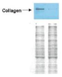
- Western blot analysis is shown using anti-Collagen I antibody to detect expression of collagen I in Wistar rat hepatic stellate cells (HSC) in control (GFP-transduced) (left lane) and PPARg-transduced cell lysates (right lane). Protein staining shown below each blot depicts equal protein loading. An equal amount of the whole cell protein (100 µg) was separated by SDS-PAGE and electroblotted to nitro-cellulose membranes. Proteins were detected by incubating the membrane with anti-Collagen I antibody at a concentration of 0.22 µg/10 ml in TBS (100 mM Tris-HCl, 0.15 M NaCl, pH 7.4) with 5% Non-fat milk. Detection occurred by incubation with a horseradish peroxidase-conjugated secondary antibody at 1 µg/10 ml.
- Validation comment
- WB

- Submitted by
- GeneTex (provider)
- Main image
- Experimental details
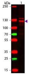
- Western blot of Human Collagen Type I. Lane 1: Human Collagen Type 1. Lane 2: None. Load: 50 ng per lane. Primary antibody: Collagen Type I antibody (GTX20292) at 1:1,000 overnight at 4°C. Secondary antibody: DyLight 649 rabbit secondary antibody at 1:20,000 for 30 min at RT. Block for 30 min at RT. Predicted/Observed size: 139 & 130 kDa, 139 & 130 kDa for Collagen Type I. Other Band(s): Collagen Type I splice variants and isoforms
- Validation comment
- WB

- Submitted by
- GeneTex (provider)
- Main image
- Experimental details
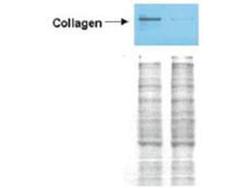
- Western blot analysis is shown using GeneTex Affinity Purified anti-Collagen I antibody (GTX20292) to detect expression of collagen I in Wistar rat hepatic stellate cells (HSC) in control (GFP-transduced) (left lane) and PPARg-transduced cell lysates (right lane). Protein staining shown below each blot depicts equal protein loading. An equal amount of the whole cell protein (100 µg) was separated by SDS-PAGE and electroblotted to nitro-cellulose membranes. Proteins were detected by incubating the membrane with anti-Collagen I antibody at a concentration of 0.22 µg/10 ml in TBS (100 mM Tris-HCl, 0.15 M NaCl, pH 7.4) with 5% Non-fat milk. Detection occurred by incubation with a horseradish peroxidase-conjugated secondary antibody at 1 µg/10 ml. Proteins were detected by a chemiluminescent method.
- Validation comment
- WB

- Submitted by
- GeneTex (provider)
- Main image
- Experimental details
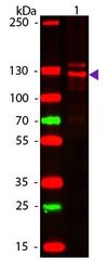
- Western blot of Human Collagen Type I. Load: 50 μg Human Collagen Type 1. Primary antibody: Collagen Type I antibody (GTX20292) at 1:1,000 overnight at 4°C. Secondary antibody: DyLight 649 rabbit secondary antibody at 1:20,000 for 30 min at RT. Block for 30 min at RT. Predicted/Observed size: 139 & 130 kDa.

Supportive validation
- Submitted by
- GeneTex (provider)
- Main image
- Experimental details
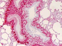
- Immunohistochemistry of Collagen I antibody (GTX20292) Tissue: human liung Fixation: formalin fixed paraffin embedded Antigen retrieval: user optimized Primary antibody: Collagen 1 1:400 Secondary antibody: Peroxidase goat anti-rabbit at 1:10,000 for 45 min at RT Localization: Strong staining was observed in the extracellular matrix of the lung. Epithelial cells were negative Staining: antibody as precipitated red signal with a hematoxylin purple nuclear counterstain

- Submitted by
- GeneTex (provider)
- Main image
- Experimental details
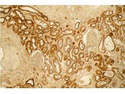
- Immunohistochemistry of Collagen I antibody (GTX20292). Tissue: Normal Kidney. Fixation: formalin fixed paraffin embedded. Antigen retrieval: No antigen retrieval was performed. Primary antibody: Collagen I 1:100 4 hours at room temperature Secondary antibody: Peroxidase goat anti-rabbit at 1:10,000 for 45 min at RT. Localization: Distal tubules in normal kidney tissue. Note the absence of staining of glomeruli. Staining: antibody as precipitated red signal with a hematoxylin purple nuclear counterstain.
