Explore
 Validate
 Learn
Learn Western blot
 ELISA
ELISAAntibody data
- Antibody Data
- Antigen structure
- References [0]
- Comments [0]
- Validations
- Western blot [2]
- Immunohistochemistry [1]
- Flow cytometry [2]
Submit
Validation data
Reference
Comment
Report error
- Product number
- GTX26577 - Provider product page

- Provider
- GeneTex
- Proper citation
- GeneTex Cat#GTX26577, RRID:AB_374559
- Product name
- Collagen I antibody (Biotin)
- Antibody type
- Polyclonal
- Reactivity
- Human, Mouse, Rat, Bovine
- Host
- Rabbit
No comments: Submit comment
Supportive validation
- Submitted by
- GeneTex (provider)
- Main image

- Experimental details
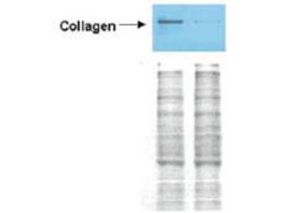
- Western Blot of Rabbit anti-Collagen I antibody (GTX26577). Lane 1: Wistar rat hepatic stellate cells (HSC) in control (GFP-transduced). Lane 2: PPARg-transduced cell lysates. Load: 100 µg per lane. Protein staining shown below each blot depicts equal protein loading. Primary antibody: anti-Collagen I antibody at 0.22 µg/10 ml for overnight at 4°C. Secondary antibody: horseradish peroxidase-conjugated rabbit secondary antibody at 1 µg/10 ml for overnight at 4°C. Block: TBS with 5% Non-fat milk. Predicted/Observed size: 138.9 kDa for Collagen I. Other band(s): none.
- Validation comment
- WB
- Submitted by
- GeneTex (provider)
- Main image

- Experimental details
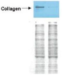
- Western Blot of Rabbit anti-Collagen I antibody (GTX26577). Lane 1: Wistar rat hepatic stellate cells (HSC) in control (GFP-transduced). Lane 2: PPARg-transduced cell lysates. Load: 100 μg per lane. Protein staining shown below each blot depicts equal protein loading. Primary antibody: anti-Collagen I antibody at 0.2¡V2 μg/10 ml for overnight at 4°C. Secondary antibody: horseradish peroxidase-conjμgated rabbit secondary antibody at 1 μg/10 ml for overnight at 4°C. Block: TBS with 5% Non-fat milk. Predicted/Observed size: 138.9 kDa for Collagen I. Other band(s): none.
Supportive validation
- Submitted by
- GeneTex (provider)
- Main image

- Experimental details
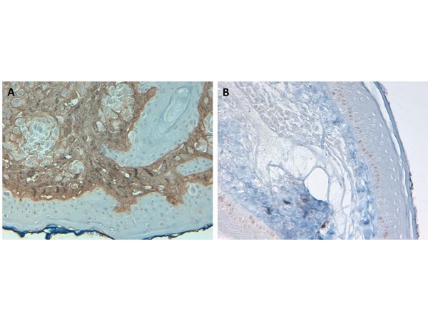
- Immunohistochemistry of Rabbit Anti-Collagen Type I Antibody (GTX26577). Tissue: Human Skin at pH9. Fixation: formalin fixed paraffin embedded. Antigen retrieval: not required. Primary antibody: Collagen Type I antibody at 10 μg/mL for 1 h at RT. Secondary antibody: Peroxidase rabbit secondary antibody at 1:10,000 for 45 min at RT. Localization: Collagen Type I is secreted in the extracellular matrix. Staining: Collagen Type I as precipitated brown signal (A) with hematoxylin purple nuclear counterstain. With corresponding negative control (B)
Supportive validation
- Submitted by
- GeneTex (provider)
- Main image
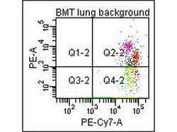
- Experimental details
- Flow Cytometry of Anti-Collagen Type I Biotin Conjμgated Antibody. Cells: mouse lung. Stimulation: none. Primary antibody: biotin conjμgated anti-collagen type I antibody. Secondary antibody: PE-conjμgated CD45 and PE-conjμgated anti-collagen type I secondary antibody.

- Submitted by
- GeneTex (provider)
- Main image
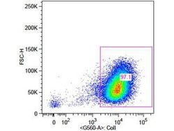
- Experimental details
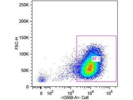
- Flow Cytometry of Rabbit Anti-Collagen 1 Antibody. Cells: primary adult human dermal fibroblast cells. Stimulation: none. Primary antibody: Biotin-Conjμgated Collagen 1 antibody at 5μg/mL for 45 min at 4°C. Secondary antibody: Rabbit Streptavidin, R-PE antibody at 1:500 for 15 min at RT.