Explore
 Validate
 Learn
Learn Western blot
Antibody data
- Antibody Data
- Antigen structure
- References [0]
- Comments [0]
- Validations
- Western blot [4]
- Immunocytochemistry [4]
- Immunohistochemistry [25]
Submit
Validation data
Reference
Comment
Report error
- Product number
- R31258 - Provider product page

- Provider
- NSJ Bioreagents
- Product name
- Collagen 1 Antibody
- Antibody type
- Polyclonal
- Description
- This highly specific Collagen 1 antibody is suitable for use in Western blot/Immunohistochemistry/Immunocytochemistry/Immunofluorescence applications with human, mouse and rat samples.
- Reactivity
- Human, Mouse, Rat
- Host
- Rabbit
- Conjugate
- Unconjugated
- Vial size
- 100 ug
- Concentration
- 0.5mg/ml if reconstituted with 0.2ml sterile DI water
- Storage
- After reconstitution, the Collagen 1 antibody can be stored for up to one month at 4oC. For long-term, aliquot and store at -20oC. Avoid repeated freezing and thawing.
No comments: Submit comment
Supportive validation
- Submitted by
- NSJ Bioreagents (provider)
- Main image
- Experimental details
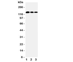
- Western blot testing of Collagen type 1 antibody and Lane 1: rat lung; 2: human placenta; 3: rat testis tissue lysate. Expected/observed size ~130KD
- Submitted by
- NSJ Bioreagents (provider)
- Main image
- Experimental details
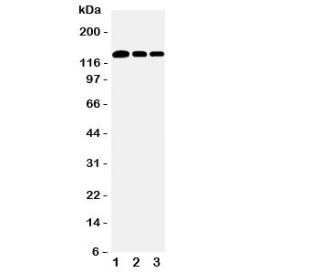
- Western blot testing of Collagen type 1 antibody and Lane 1: rat lung; 2: human placenta; 3: rat testis tissue lysate. Expected molecular weight: 140-210 kDa (precusor), 70-90 kDa (mature).

- Submitted by
- NSJ Bioreagents (provider)
- Main image

- Experimental details
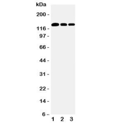
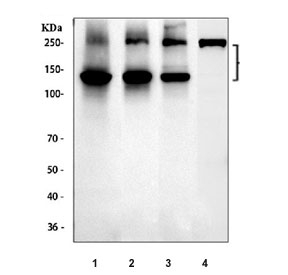
- Western blot testing of 1) human placenta, 2) rat skin, 3) mouse skin and 4) mouse NIH 3T3 cell lysate with Collagen 1 antibody. Expected molecular weight: 140-210 kDa (precusor), 70-90 kDa (mature).
- Submitted by
- NSJ Bioreagents (provider)
- Main image

- Experimental details
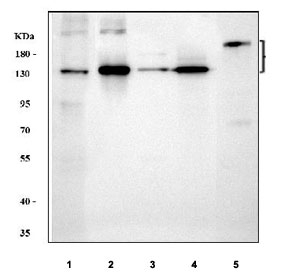
- Western blot testing of 1) human placenta, 2) rat skin, 3) rat lung, 4) mouse skin and 5) mouse NIH 3T3 cell lysate with Collagen 1 antibody. Expected molecular weight: 140-210 kDa (precusor), 70-90 kDa (mature).
Supportive validation
- Submitted by
- NSJ Bioreagents (provider)
- Main image
- Experimental details
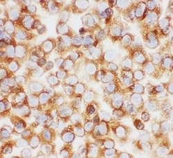
- ICC testing of Collagen type 1 antibody and NIH3T3 cells

- Submitted by
- NSJ Bioreagents (provider)
- Main image
- Experimental details
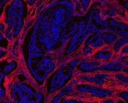
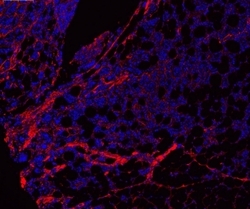
- Immunofluorescent staining of FFPE human breast tissue with Collagen 1 antibody (red) and DAPI counterstain (blue). HIER: boil tissue sections in pH6, 10mM citrate buffer, for 10-20 min and allow to cool before testing.
- Submitted by
- NSJ Bioreagents (provider)
- Main image
- Experimental details
- Immunofluorescent staining of FFPE human tonsil tissue with Collagen 1 antibody (red) and DAPI counterstain (blue). HIER: boil tissue sections in pH6, 10mM citrate buffer, for 10-20 min and allow to cool before testing.

- Submitted by
- NSJ Bioreagents (provider)
- Main image

- Experimental details
- ICC testing of Collagen 1 antibody and NIH3T3 cells. HIER: boil tissue sections in pH6, 10mM citrate buffer, for 10-20 min and allow to cool before testing.

Supportive validation
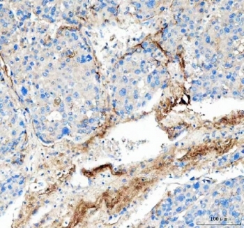
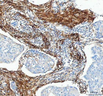
- Submitted by
- NSJ Bioreagents (provider)
- Main image
- Experimental details
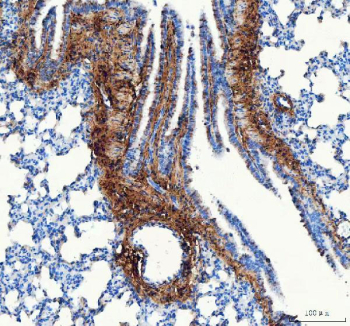
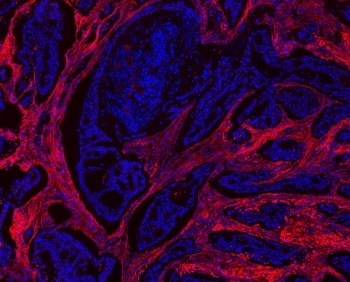
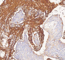
- IHC-P: Collagen 1 antibody testing of human intestine cancer tissue. HIER: boil tissue sections in pH6, 10mM citrate buffer, for 10-20 min and allow to cool before testing.

- Submitted by
- NSJ Bioreagents (provider)
- Main image
- Experimental details
- IHC-F testing of Collagen type 1 antibody and human placenta tissue

- Submitted by
- NSJ Bioreagents (provider)
- Main image
- Experimental details
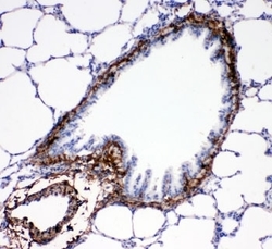
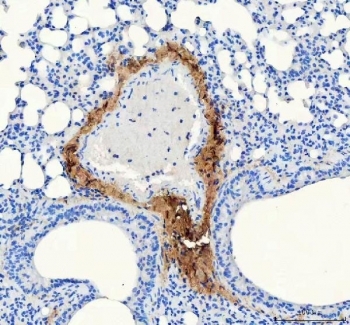
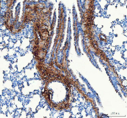
- IHC-P: Collagen type 1 antibody testing of rat lung tissue

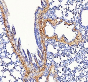
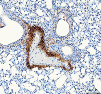
- Submitted by
- NSJ Bioreagents (provider)
- Main image

- Experimental details
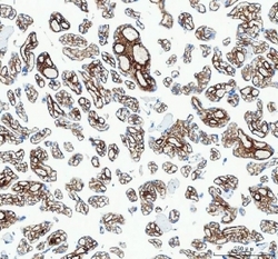
- IHC-P: Collagen 1 antibody testing of mouse lung tissue at 2ug/ml. HIER: boil tissue sections in pH6, 10mM citrate buffer, for 10-20 min and allow to cool before testing.
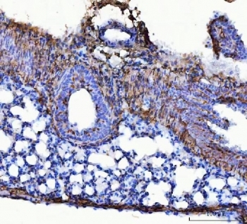
- Submitted by
- NSJ Bioreagents (provider)
- Main image
- Experimental details
- IHC staining of FFPE human placental tissue with Collagen 1 antibody. HIER: boil tissue sections in pH8 EDTA for 20 min and allow to cool before testing.
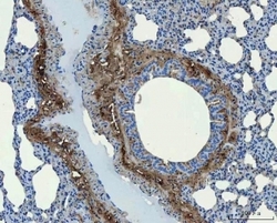
- Submitted by
- NSJ Bioreagents (provider)
- Main image

- Experimental details
- IHC-P: Collagen 1 antibody testing of rat lung tissue. HIER: boil tissue sections in pH6, 10mM citrate buffer, for 10-20 min and allow to cool before testing.

- Submitted by
- NSJ Bioreagents (provider)
- Main image

- Experimental details
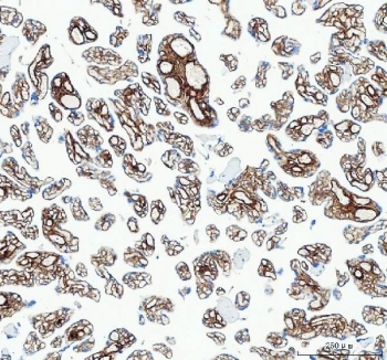
- IHC staining of FFPE mouse lung tissue with Collagen 1 antibody. HIER: boil tissue sections in pH8 EDTA for 20 min and allow to cool before testing.
- Submitted by
- NSJ Bioreagents (provider)
- Main image

- Experimental details
- IHC-P: Collagen 1 antibody testing of mouse lung tissue at 2ug/ml. HIER: boil tissue sections in pH6, 10mM citrate buffer, for 10-20 min and allow to cool before testing.
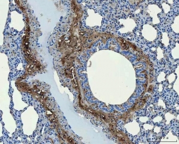
- Submitted by
- NSJ Bioreagents (provider)
- Main image
- Experimental details
- IHC-P: Collagen 1 antibody testing of rat lung tissue at 2ug/ml. HIER: boil tissue sections in pH6, 10mM citrate buffer, for 10-20 min and allow to cool before testing.
- Submitted by
- NSJ Bioreagents (provider)
- Main image

- Experimental details
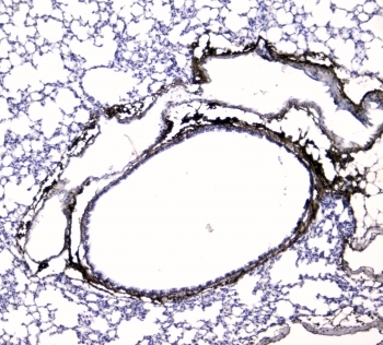
- IHC staining of FFPE rat lung tissue with Collagen 1 antibody. HIER: boil tissue sections in pH8 EDTA for 20 min and allow to cool before testing.
- Submitted by
- NSJ Bioreagents (provider)
- Main image

- Experimental details
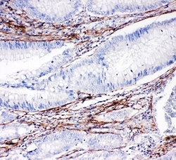
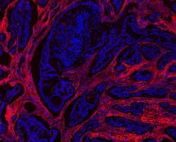
- IHC staining of FFPE human esophageal squamous carcinoma tissue with Collagen 1 antibody. HIER: boil tissue sections in pH8 EDTA for 20 min and allow to cool before testing.

- Submitted by
- NSJ Bioreagents (provider)
- Main image

- Experimental details
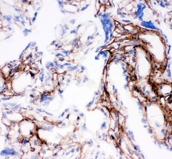
- IHC staining of FFPE human liver cancer tissue with Collagen 1 antibody. HIER: boil tissue sections in pH8 EDTA for 20 min and allow to cool before testing.
- Submitted by
- NSJ Bioreagents (provider)
- Main image
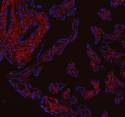
- Experimental details
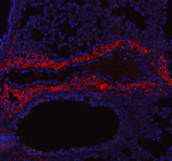
- Immunofluorescent staining of FFPE human placental tissue with Collagen 1 antibody (red) and DAPI counterstain (blue). HIER: boil tissue sections in pH8 EDTA buffer, for 10-20 min and allow to cool before testing.
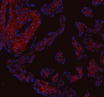
- Submitted by
- NSJ Bioreagents (provider)
- Main image

- Experimental details
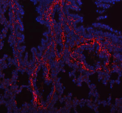
- Immunofluorescent staining of FFPE mouse lung tissue with Collagen 1 antibody (red) and DAPI counterstain (blue). HIER: boil tissue sections in pH8 EDTA buffer, for 10-20 min and allow to cool before testing.
- Submitted by
- NSJ Bioreagents (provider)
- Main image
- Experimental details
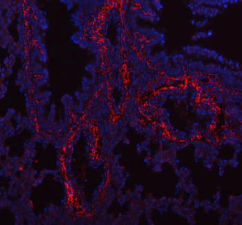
- Immunofluorescent staining of FFPE rat lung tissue with Collagen 1 antibody (red) and DAPI counterstain (blue). HIER: boil tissue sections in pH8 EDTA buffer, for 10-20 min and allow to cool before testing.
- Submitted by
- NSJ Bioreagents (provider)
- Main image
- Experimental details
- Immunofluorescent staining of FFPE human breast tissue with Collagen 1 antibody (red) and DAPI counterstain (blue). HIER: boil tissue sections in pH6 citrate buffer for 10-20 min and allow to cool before testing.

- Submitted by
- NSJ Bioreagents (provider)
- Main image

- Experimental details
- Immunofluorescent staining of FFPE human tonsil tissue with Collagen 1 antibody (red) and DAPI counterstain (blue). HIER: boil tissue sections in pH6 citrate buffer for 10-20 min and allow to cool before testing.

- Submitted by
- NSJ Bioreagents (provider)
- Main image
- Experimental details
- IHC staining of FFPE human liver cancer tissue with Collagen 1 antibody, HRP secondary and DAB substrate. HIER: boil tissue sections in pH8 EDTA for 20 min and allow to cool before testing.

- Submitted by
- NSJ Bioreagents (provider)
- Main image

- Experimental details
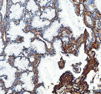
- IHC staining of FFPE human lung adenocarcinoma tissue with Collagen 1 antibody, HRP secondary and DAB substrate. HIER: boil tissue sections in pH8 EDTA for 20 min and allow to cool before testing.
- Submitted by
- NSJ Bioreagents (provider)
- Main image
- Experimental details
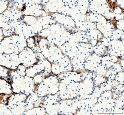
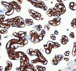
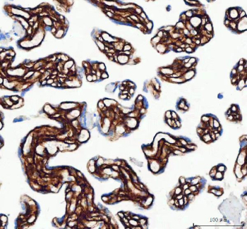
- IHC staining of FFPE human renal cell carcinoma tissue with Collagen 1 antibody, HRP secondary and DAB substrate. HIER: boil tissue sections in pH8 EDTA for 20 min and allow to cool before testing.

- Submitted by
- NSJ Bioreagents (provider)
- Main image

- Experimental details
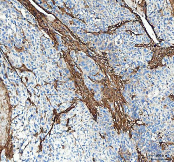
- IHC staining of FFPE human bladder urothelial carcinoma tissue with Collagen 1 antibody, HRP secondary and DAB substrate. HIER: boil tissue sections in pH8 EDTA for 20 min and allow to cool before testing.
- Submitted by
- NSJ Bioreagents (provider)
- Main image
- Experimental details
- IHC staining of FFPE human placental tissue with Collagen 1 antibody, HRP secondary and DAB substrate. HIER: boil tissue sections in pH8 EDTA for 20 min and allow to cool before testing.
- Submitted by
- NSJ Bioreagents (provider)
- Main image

- Experimental details
- IHC staining of FFPE human esophageal squamous carcinoma tissue with Collagen 1 antibody, HRP secondary and DAB substrate. HIER: boil tissue sections in pH8 EDTA for 20 min and allow to cool before testing.
- Submitted by
- NSJ Bioreagents (provider)
- Main image

- Experimental details
- IHC staining of FFPE mouse lung tissue with Collagen 1 antibody, HRP secondary and DAB substrate. HIER: boil tissue sections in pH8 EDTA for 20 min and allow to cool before testing.
- Submitted by
- NSJ Bioreagents (provider)
- Main image
- Experimental details
- IHC staining of FFPE rat lung tissue with Collagen 1 antibody, HRP secondary and DAB substrate. HIER: boil tissue sections in pH8 EDTA for 20 min and allow to cool before testing.