Explore
 Validate
 Learn
Learn Western blot
 ELISA
ELISAAntibody data
- Antibody Data
- Antigen structure
- References [0]
- Comments [0]
- Validations
- Western blot [5]
- Immunohistochemistry [11]
Submit
Validation data
Reference
Comment
Report error
- Product number
- NB600-408-0.1mg - Provider product page

- Provider
- Novus Biologicals
- Product name
- Rabbit Polyclonal Collagen I Antibody
- Antibody type
- Polyclonal
- Description
- Immunogen affinity purified. This Collagen I antibody has typically less than 1% cross-reactivity against other types of collagens was detected by ELISA against purified standards. Some class-specific anti-collagens may be specific for three-dimensional epitopes which may result in diminished reactivity with denatured collagen or formalin-fixed, paraffin embedded tissues. This antibody reacts with most mammalian Type I collagens and has negligible cross-reactivity with Type II, III, IV, V or VI collagens. Non-specific cross-reaction of anti-collagen antibodies with other human serum proteins or non-collagen extracellular matrix proteins is negligible.
- Reactivity
- Human, Mouse, Rat, Bovine, Feline, Porcine, Rabbit
- Host
- Rabbit
- Isotype
- IgG
- Vial size
- 0.1 mg
- Concentration
- 1.0 mg/ml
- Storage
- Store at -20 degreesC.
No comments: Submit comment
Supportive validation
- Submitted by
- Novus Biologicals (provider)
- Main image

- Experimental details
- Western Blot: Collagen I Antibody [NB600-408] - Cell lysate from human trabecular meshwork. Antibody at 1:1.000. WB image submitted by a verified customer review.

- Submitted by
- Novus Biologicals (provider)
- Main image

- Experimental details
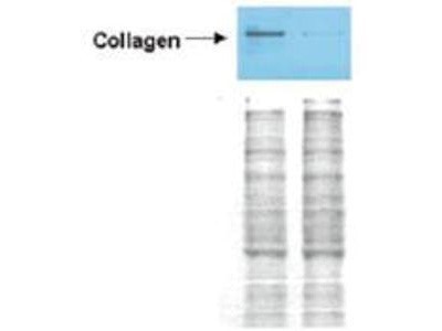
- Western Blot: Collagen I Antibody [NB600-408] - Detection of collagen I in Wistar rat hepatic stellate cells (HSC) in control (GFP-transduced) (left lane) and PPARg-transduced cell lysates (right lane). Protein staining shown below each blot depicts equal protein loading. An equal amount of the whole cell protein (100 ug) was separated by SDS-PAGE and electroblotted to nitro-cellulose membranes. Proteins were detected by incubating the membrane with anti-Collagen I antibody at a concentration of 0.2-2 ug/10 mL in TBS (100 mM Tris-HCl, 0.15 M NaCl, pH 7.4) with 5% non-fat milk. Detection occurred by incubation with a HRP conjugated secondary antibody at 1 ug/10 mL. Proteins were detected by a chemiluminescent method using the PIERCE ECL kit (Amersham Biosciences).
- Submitted by
- Novus Biologicals (provider)
- Main image
- Experimental details
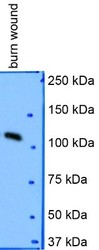
- Western Blot: Collagen I Antibody [NB600-408] - Analysis in porcine burn wound lysate using anti-Collagen I alpha 1 antibody. WB image submitted by a verified customer review.

- Submitted by
- Novus Biologicals (provider)
- Main image
- Experimental details
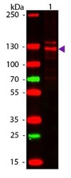
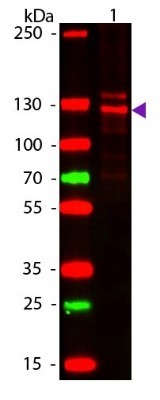
- Western Blot: Collagen I Antibody [NB600-408] - Lane 1: Human Collagen Type 1. Lane 2: None. Load: 50 ng per lane. Primary antibody: Collagen Type I antibody at 1:1000 overnight at 4C. Secondary antibody: DyLight 649 rabbit secondary antibody at 1:20000 for 30 min at RT. Block: incubated with blocking buffer for 30 min at RT. Predicted/Observed size: 139 & 130 kDa, 139 & 130 kDa for Collagen Type I. Other Band(s): Collagen Type I splice variants and isoforms.
- Submitted by
- Novus Biologicals (provider)
- Main image

- Experimental details
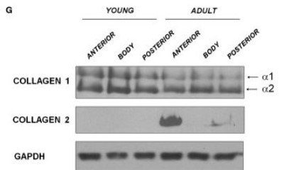
- Western Blot: Collagen I Antibody [NB600-408] - Representative Western blot image for Collagen I, Collagen 2 and GAPDH. N/group = 8. Image collected and cropped by CiteAb from the following publication (http://doi.wiley.com/10.1111/jcmm.12367) licensed under a CC-BY licence.
Supportive validation
- Submitted by
- Novus Biologicals (provider)
- Main image

- Experimental details
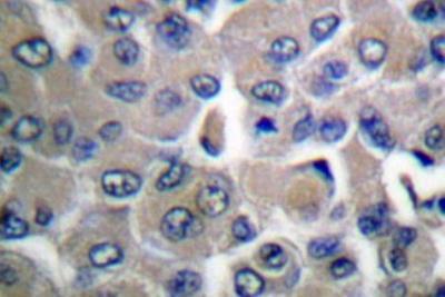
- Immunohistochemistry-Paraffin: Collagen I Antibody [NB600-408] - Balb/3T3 mouse embryonic fibroblast cell line stained with Collagen I antibody. IHC-P image submitted by a verified customer review.
- Submitted by
- Novus Biologicals (provider)
- Main image
- Experimental details
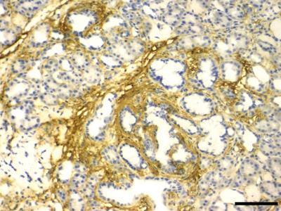
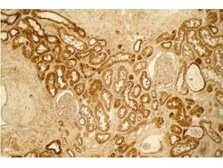
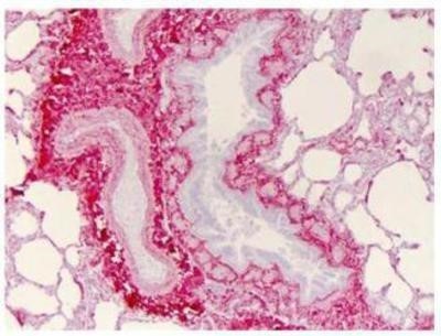
- Immunohistochemistry: Collagen I Antibody [NB600-408] - Analysis of FFPE human lung. Primary antibody: Collagen I 1:400. Secondary antibody: Peroxidase goat anti-rabbit at 1:10,000 for 45 min at RT. Localization: Strong staining was observed in the extracellular matrix of the lung. Epithelial cells were negative. Staining: antibody as precipitated red signal with a hematoxylin purple nuclear counterstain.
- Submitted by
- Novus Biologicals (provider)
- Main image
- Experimental details
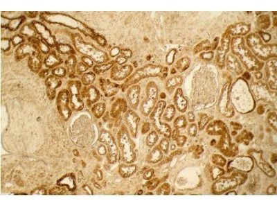
- Immunohistochemistry: Collagen I Antibody [NB600-408] - Immunohistological staining of EG treated rat kidneys for Collagen. Magnification X40, Scale bar 100 um. Image collected and cropped by CiteAb from the following publication (http://dx.plos.org/10.1371/journal.pone.0185009), licensed under a CC-BY licence.

- Submitted by
- Novus Biologicals (provider)
- Main image
- Experimental details
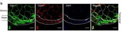
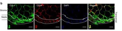
- Immunohistochemistry: Collagen I Antibody [NB600-408] - Macro- and micro-architecture of human adipose tissue lobule. Representative immunostaining of the human AT lobule with Collagen I (COL1), CD34, and DAPI. The position of the septa is underlined, scale bar: 100 um. Image collected and cropped by CiteAb from the following publication (http://www.nature.com/articles/s41467-019-09992-3) licensed under a CC-BY licence.
- Submitted by
- Novus Biologicals (provider)
- Main image
- Experimental details
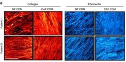
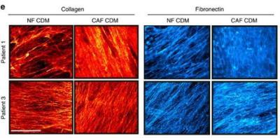
- Immunohistochemistry: Collagen I Antibody [NB600-408] - Patient-derived CAF and NF CDMs are architecturally and functionally distinct. Representative western blots showing Collagen I (red) and fibronectin (blue) staining of patient #1 and #3 NF and CAF CDM. Scale bar, 20 um. (f) Representative SEM images of TIFF, Patient #1 NF and CAF CDM. Scale bar, 5 um. Image collected and cropped by CiteAb from the following publication (http://www.nature.com/doifinder/10.1038/ncomms12237) licensed under a CC-BY licence.
- Submitted by
- Novus Biologicals (provider)
- Main image

- Experimental details
- Immunohistochemistry-Paraffin: Collagen I Antibody [NB600-408] - Human lung tissue section stained with Collagen I antibody at 1:400.
- Submitted by
- Novus Biologicals (provider)
- Main image
- Experimental details
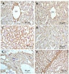
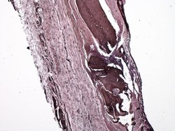
- Immunohistochemistry-Paraffin: Collagen I Antibody [NB600-408] - FFPE right lobe of the liver tissue section. A:Central Vein (CV) fibrosis, B: Non-fibrotic CV, C: Perisinusodial fibrosis, D: Non-fibrotic area, E: Protat tract fibrosis, F: Septal fibrosis (arrow). Antigen retrieval: not required. Primary antibody: Anti-collagen type I at 1:1250 for 4C for 24hr. Secondary antibody: Peroxidase biotin-streptavidin rabbit secondary antibody at 1:10,000 for 45 min at RT. Localization: Anti-collagen type I is intra and extracellular. Staining: 3.3'-diaminobenzidine tetrahydrochloride was used as the chromogen. Nuclei were counterstained purple with hematoxylin.

- Submitted by
- Novus Biologicals (provider)
- Main image
- Experimental details
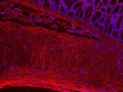
- Immunohistochemistry-Paraffin: Collagen I Antibody [NB600-408] - Rat colon tissue stained with Collagen I antibody (red) and Hoechst (blue). IHC-P image submitted by a verified customer review.

- Submitted by
- Novus Biologicals (provider)
- Main image
- Experimental details
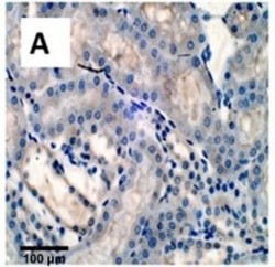
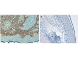
- Immunohistochemistry-Paraffin: Collagen I Antibody [NB600-408] - Analysis of HRP conjugate of NB600-408. FFPE Human Skin tissue at pH9. Primary antibody: Collagen Type I antibody at 10 ug/mL for 1 h at RT. Localization: Collagen Type I is secreted in the extracellular matrix. Staining: Collagen Type I as precipitated brown signal (A) with hematoxylin purple nuclear counterstain. With corresponding negative conrol (B).

- Submitted by
- Novus Biologicals (provider)
- Main image
- Experimental details
- Immunohistochemistry-Paraffin: Collagen I Antibody [NB600-408] - Imaging of rat calvarial defect bone. IHC-P image submitted by a verified customer review.

- Submitted by
- Novus Biologicals (provider)
- Main image

- Experimental details
- Immunohistochemistry-Paraffin: Collagen I Antibody [NB600-408] - Mouse pancreas tissue stained with Collagen I antibody. IHC-P image submitted by a verified customer review.