 Explore
 Validate
 Learn
Learn Western blot
 ELISA
ELISAAntibody data
- Antibody Data
- Antigen structure
- References [0]
- Comments [0]
- Validations
- Western blot [2]
- Immunohistochemistry [3]
Submit
Validation data
Reference
Comment
Report error
- Product number
- LS-C745280 - Provider product page

- Provider
- LSBio
- Product name
- COL1A1 / Collagen I Alpha 1 Antibody LS-C745280
- Antibody type
- Polyclonal
- Reactivity
- Human, Bovine
- Host
- Rabbit
- Isotype
- IgG
- Storage
- Store vial at -20°C or below prior to opening. Dilute 1:10 to minimize loss. Store the vial at -20°C or below after dilution. Avoid freeze-thaw cycles.
No comments: Submit comment
Supportive validation
- Submitted by
- LSBio (provider)
- Main image
- Experimental details
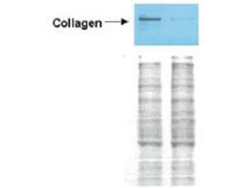
- Western blot analysis is shown using the Affinity Purified anti-Collagen I antibody to detect expression of collagen I in Wistar rat hepatic stellate cells (HSC) in control (GFP-transduced) (left lane) and PPARg-transduced cell lysates (right lane). Protein staining shown below each blot depicts equal protein loading. An equal amount of the whole cell protein (100 µg) was separated by SDS-PAGE and electroblotted to nitro-cellulose membranes. Proteins were detected by incubating the membrane with anti-Collagen I antibody at a concentration of 0.2–2 µg/10 ml in TBS (100 mM Tris-HCl, 0.15 M NaCl, pH 7.4) with 5% Non-fat milk. Detection occurred by incubation with a horseradish peroxidase-conjugated secondary antibody at 1 µg/10 ml. Proteins were detected by a chemiluminescent method using the PIERCE ECL kit (Amersham Biosciences). Other detection systems will yield similar results. See Hazra et al. (2004) for additional details.

- Submitted by
- LSBio (provider)
- Main image

- Experimental details
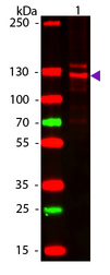
- Western blot of Human Collagen Type I. Lane 1: Human Collagen Type 1. Load: 50 ng per lane. Primary antibody: Collagen Type I antibody at 1:1,000 overnight at 4°C. Secondary antibody: DyLight 649 rabbit secondary antibody at 1:20,000 for 30 min at RT. Block: MB-070 for 30 min at RT. Predicted/Observed size: 139 & 130 kDa, 139 & 130 kDa for Collagen Type I. Other Band(s): Collagen Type I splice variants and isoforms.
Supportive validation
- Submitted by
- LSBio (provider)
- Main image

- Experimental details
- Immunohistochemistry of Collagen I antibody. Tissue: human lung. Fixation: formalin fixed paraffin embedded. Antigen retrieval: user optimized. Primary antibody: Collagen 1 at 1:400. Secondary antibody: Peroxidase goat anti-rabbit at 1:10,000 for 45 min at RT. Localization: Strong staining was observed in the extracellular matrix of the lung. Epithelial cells were negative. Staining: antibody as precipitated red signal with a hematoxylin purple nuclear counterstain.

- Submitted by
- LSBio (provider)
- Main image

- Experimental details
- Affinity Purified anti-Collagen I antibody was used at a 1:100 dilution to detect distal tubules in normal kidney tissue. Note the absence of staining of glomeruli. The antibody was reacted with antibody for 4 hours at room temperature followed by the addition of secondary antibody and substrate reaction. Tissue was formalin-fixed and paraffin embedded. No antigen retrieval was performed.

- Submitted by
- LSBio (provider)
- Main image

- Experimental details
- Immunohistochemistry of rabbit Anti-collagen type I antibody. Tissue: right lobe of the liver section. A:Central Vein (CV) fibrosis, B: Non-fibrotic CV, C: perisinusoidal fibrosis, D: Non-fibrotic area, E: Portal tract fibrosis, F: Septal fibrosis (arrow). Fixation: formalin fixed paraffin embedded. Antigen retrieval: not required. Primary antibody: Anti-collagen type I at 1:1250 for 4°C for 24hr. Secondary antibody: Peroxidase biotin-streptavidin rabbit secondary antibody at 1:10,000 for 45 min at RT. Localization: Anti-collagen type I is intra and extracellular. Staining: 3.3’-diaminobenzidine tetrahydrochloride was used as the chromogen. Nuclei were counterstained purple with hematoxylin.
