 Explore
 Validate
 Learn
Learn Western blot
 Immunocytochemistry
Antibody data
- Antibody Data
- Antigen structure
- References [9]
- Comments [0]
- Validations
- Immunocytochemistry [6]
- Immunoprecipitation [1]
- Immunohistochemistry [5]
- Other assay [4]
Submit
Validation data
Reference
Comment
Report error
- Product number
- PA5-29569 - Provider product page

- Provider
- Invitrogen Antibodies
- Product name
- COL1A1 Polyclonal Antibody
- Antibody type
- Polyclonal
- Antigen
- Recombinant full-length protein
- Description
- Recommended positive controls: SK-N-SH, SK-N-AS, NIH3T3. Predicted reactivity: Mouse (96%), Rat (94%), Xenopus laevis (83%), Dog (98%), Bovine (98%). Store product as a concentrated solution. Centrifuge briefly prior to opening the vial.
- Reactivity
- Human, Mouse, Rat
- Host
- Rabbit
- Isotype
- IgG
- Vial size
- 100 μL
- Concentration
- 0.09 mg/mL
- Storage
- Store at 4°C short term. For long term storage, store at -20°C, avoiding freeze/thaw cycles.
Submitted references An Injectable Fibrin Scaffold Rich in Growth Factors for Skin Repair.
Deficient Endoplasmic Reticulum Acetyl-CoA Import in Pancreatic Acinar Cells Leads to Chronic Pancreatitis.
Impaired non-canonical transforming growth factor-β signalling prevents profibrotic phenotypes in cultured peptidylarginine deiminase 4-deficient murine cardiac fibroblasts.
Dystrophin Deficiency Causes Progressive Depletion of Cardiovascular Progenitor Cells in the Heart.
Translation of Tudor-SN, a novel terminal oligo-pyrimidine (TOP) mRNA, is regulated by the mTORC1 pathway in cardiomyocytes.
Small molecule inhibitor of HSP47 prevents pro-fibrotic mechanisms of fibroblasts in vitro.
FOXM1 is a critical driver of lung fibroblast activation and fibrogenesis.
Sirtuin1 promotes osteogenic differentiation through downregulation of peroxisome proliferator-activated receptor γ in MC3T3-E1 cells.
MiR-132 regulates osteogenic differentiation via downregulating Sirtuin1 in a peroxisome proliferator-activated receptor β/δ-dependent manner.
Shao Z, Lyu C, Teng L, Xie X, Sun J, Zou D, Lu J
BioMed research international 2021;2021:8094932
BioMed research international 2021;2021:8094932
Deficient Endoplasmic Reticulum Acetyl-CoA Import in Pancreatic Acinar Cells Leads to Chronic Pancreatitis.
Cooley MM, Thomas DDH, Deans K, Peng Y, Lugea A, Pandol SJ, Puglielli L, Groblewski GE
Cellular and molecular gastroenterology and hepatology 2021;11(3):725-738
Cellular and molecular gastroenterology and hepatology 2021;11(3):725-738
Impaired non-canonical transforming growth factor-β signalling prevents profibrotic phenotypes in cultured peptidylarginine deiminase 4-deficient murine cardiac fibroblasts.
Akboua H, Eghbalzadeh K, Keser U, Wahlers T, Paunel-Görgülü A
Journal of cellular and molecular medicine 2021 Oct;25(20):9674-9684
Journal of cellular and molecular medicine 2021 Oct;25(20):9674-9684
Dystrophin Deficiency Causes Progressive Depletion of Cardiovascular Progenitor Cells in the Heart.
Jelinkova S, Sleiman Y, Fojtík P, Aimond F, Finan A, Hugon G, Scheuermann V, Beckerová D, Cazorla O, Vincenti M, Amedro P, Richard S, Jaros J, Dvorak P, Lacampagne A, Carnac G, Rotrekl V, Meli AC
International journal of molecular sciences 2021 May 10;22(9)
International journal of molecular sciences 2021 May 10;22(9)
Translation of Tudor-SN, a novel terminal oligo-pyrimidine (TOP) mRNA, is regulated by the mTORC1 pathway in cardiomyocytes.
Gan S, Su C, Ma J, Liu M, Cui X, Xin L, Ren Y, Gao X, Ge L, Wei M, Yang J
RNA biology 2021 Jun;18(6):900-913
RNA biology 2021 Jun;18(6):900-913
Small molecule inhibitor of HSP47 prevents pro-fibrotic mechanisms of fibroblasts in vitro.
Miyamura T, Sakamoto N, Kakugawa T, Taniguchi H, Akiyama Y, Okuno D, Moriyama S, Hara A, Kido T, Ishimoto H, Yamaguchi H, Miyazaki T, Obase Y, Ishimatsu Y, Tanaka Y, Mukae H
Biochemical and biophysical research communications 2020 Sep 24;530(3):561-565
Biochemical and biophysical research communications 2020 Sep 24;530(3):561-565
FOXM1 is a critical driver of lung fibroblast activation and fibrogenesis.
Penke LR, Speth JM, Dommeti VL, White ES, Bergin IL, Peters-Golden M
The Journal of clinical investigation 2018 Jun 1;128(6):2389-2405
The Journal of clinical investigation 2018 Jun 1;128(6):2389-2405
Sirtuin1 promotes osteogenic differentiation through downregulation of peroxisome proliferator-activated receptor γ in MC3T3-E1 cells.
Qu B, Ma Y, Yan M, Gong K, Liang F, Deng S, Jiang K, Ma Z, Pan X
Biochemical and biophysical research communications 2016 Sep 9;478(1):439-445
Biochemical and biophysical research communications 2016 Sep 9;478(1):439-445
MiR-132 regulates osteogenic differentiation via downregulating Sirtuin1 in a peroxisome proliferator-activated receptor β/δ-dependent manner.
Gong K, Qu B, Liao D, Liu D, Wang C, Zhou J, Pan X
Biochemical and biophysical research communications 2016 Sep 9;478(1):260-267
Biochemical and biophysical research communications 2016 Sep 9;478(1):260-267
No comments: Submit comment
Supportive validation
- Submitted by
- Invitrogen Antibodies (provider)
- Main image

- Experimental details
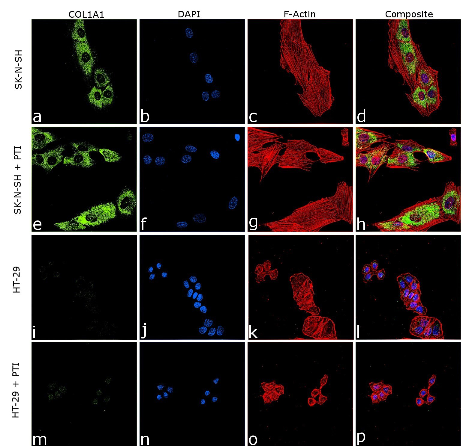
- Immunofluorescence analysis of COL1A1 was performed using 70% confluent log phase SK-N-SH and HT-29 cells. The cells were fixed with 4% paraformaldehyde for 5 minutes, permeabilized with 0.1% Triton™ X-100 for 5 minutes, and blocked with 2% BSA for 45 minutes at room temperature. The cells were labeled with COL1A1 Polyclonal Antibody (Product # PA5-29569) at 1:100 dilution in 0.1% BSA, incubated at 4 degree celsius overnight and then labeled with Donkey anti-Rabbit IgG (H+L) Highly Cross-Adsorbed Secondary Antibody, Alexa Fluor Plus 488 (Product # A32790), (1:2000 dilution), for 45 minutes at room temperature (Panel a: Green). Nuclei (Panel b:Blue) were stained with ProLong™ Diamond Antifade Mountant with DAPI (Product # P36962). F-actin (Panel c: Red) was stained with Rhodamine Phalloidin (Product # R415, 1:300). Panel d represents the merged image showing cytosolic localization. Panel (a-d) shows representative SK-N-SH control cell, whereas Panel (e-h) represent SK-N-SH with PTI treatment. Similarly, panel (i-l) represent HT-29 control cells and Panel (m-p) represent HT-29 with PTI treatment. The images were captured at 60 magnification.

- Submitted by
- Invitrogen Antibodies (provider)
- Main image

- Experimental details
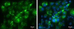
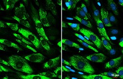
- Immunocytochemistry-Immunofluorescence analysis of COL1A1 was performed in SK-N-AS cells fixed in 4% paraformaldehyde at RT for 15 min. Green: COL1A1 Polyclonal Antibody (Product # PA5-29569) diluted at 1:500. Blue: Hoechst 33342 staining. Scale bar = 10 µm.

- Submitted by
- Invitrogen Antibodies (provider)
- Main image

- Experimental details
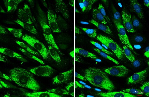
- COL1A1 Polyclonal Antibody detects COL1A1 protein at cytoplasm by immunofluorescent analysis. Sample: SK-N-SH cells were fixed in 4% paraformaldehyde at RT for 15 min. Green: COL1A1 stained by COL1A1 Polyclonal Antibody (Product # PA5-29569) diluted at 1:500. Blue: Fluoroshield with DAPI . Scale bar= 10 µm.

- Submitted by
- Invitrogen Antibodies (provider)
- Main image
- Experimental details
- Immunocytochemistry-Immunofluorescence analysis of COL1A1 was performed in SK-N-AS cells fixed in 4% paraformaldehyde at RT for 15 min. Green: COL1A1 Polyclonal Antibody (Product # PA5-29569) diluted at 1:500. Blue: Hoechst 33342 staining. Scale bar = 10 µm.

- Submitted by
- Invitrogen Antibodies (provider)
- Main image

- Experimental details
- Immunofluorescence analysis of COL1A1 was performed using 70% confluent log phase SK-N-SH and HT-29 cells. The cells were fixed with 4% paraformaldehyde for 5 minutes, permeabilized with 0.1% Triton™ X-100 for 5 minutes, and blocked with 2% BSA for 45 minutes at room temperature. The cells were labeled with COL1A1 Polyclonal Antibody (Product # PA5-29569) at 1:100 dilution in 0.1% BSA, incubated at 4 degree celsius overnight and then labeled with Donkey anti-Rabbit IgG (H+L) Highly Cross-Adsorbed Secondary Antibody, Alexa Fluor Plus 488 (Product # A32790), (1:2000 dilution), for 45 minutes at room temperature (Panel a: Green). Nuclei (Panel b:Blue) were stained with ProLong™ Diamond Antifade Mountant with DAPI (Product # P36962). F-actin (Panel c: Red) was stained with Rhodamine Phalloidin (Product # R415, 1:300). Panel d represents the merged image showing cytosolic localization. Panel (a-d) shows representative SK-N-SH control cell, whereas Panel (e-h) represent SK-N-SH with PTI treatment. Similarly, panel (i-l) represent HT-29 control cells and Panel (m-p) represent HT-29 with PTI treatment. The images were captured at 60 magnification.
- Submitted by
- Invitrogen Antibodies (provider)
- Main image
- Experimental details
- COL1A1 Polyclonal Antibody detects COL1A1 protein at cytoplasm by immunofluorescent analysis. Sample: SK-N-SH cells were fixed in 4% paraformaldehyde at RT for 15 min. Green: COL1A1 stained by COL1A1 Polyclonal Antibody (Product # PA5-29569) diluted at 1:500. Blue: Fluoroshield with DAPI . Scale bar= 10 µm.
Supportive validation
- Submitted by
- Invitrogen Antibodies (provider)
- Main image

- Experimental details
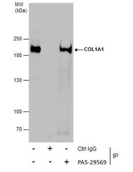
- Immunoprecipitation of COL1A1 was performed in SK-N-AS whole cell extracts using 5 µg of COL1A1 Polyclonal Antibody (Product # PA5-29569). Samples were transferred to a membrane and probed with COL1A1 Polyclonal Antibody as a primary antibody and an HRP-conjugated anti-Rabbit IgG was used as a secondary antibody.

Supportive validation
- Submitted by
- Invitrogen Antibodies (provider)
- Main image
- Experimental details
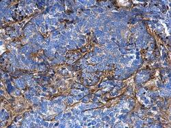
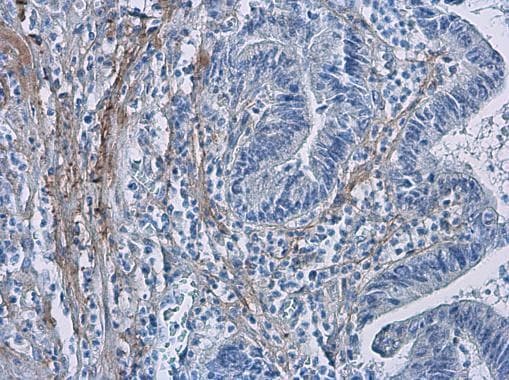
- COL1A1 Polyclonal Antibody detects COL1A1 protein at secreted on human endometrial carcinoma by immunohistochemical analysis. Sample: Paraffin-embedded human endometrial carcinoma. COL1A1 Polyclonal Antibody (Product # PA5-29569) diluted at 1:500. Antigen Retrieval: EDTA based buffer, pH 8.0, 15 min.

- Submitted by
- Invitrogen Antibodies (provider)
- Main image

- Experimental details
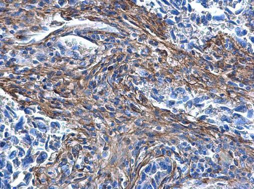
- COL1A1 Polyclonal Antibody detects COL1A1 protein at secreted on human lung carcinoma by immunohistochemical analysis. Sample: Paraffin-embedded human lung carcinoma. COL1A1 Polyclonal Antibody (Product # PA5-29569) diluted at 1:500. Antigen Retrieval: EDTA based buffer, pH 8.0, 15 min.
- Submitted by
- Invitrogen Antibodies (provider)
- Main image

- Experimental details
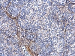
- Immunohistochemistry (Paraffin) analysis of COL1A1 was performed in paraffin-embedded human breast carcinoma tissue using COL1A1 Polyclonal Antibody (Product # PA5-29569) at a dilution of 1:500.
- Submitted by
- Invitrogen Antibodies (provider)
- Main image
- Experimental details
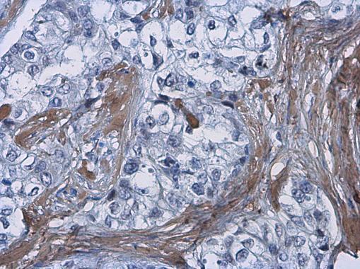
- COL1A1 Polyclonal Antibody detects COL1A1 protein at secreted on human cervical carcinoma by immunohistochemical analysis. Sample: Paraffin-embedded human cervical carcinoma. COL1A1 Polyclonal Antibody (Product # PA5-29569) dilution: 1:500. Antigen Retrieval: EDTA based buffer, pH 8.0, 15 min.

- Submitted by
- Invitrogen Antibodies (provider)
- Main image

- Experimental details
- Immunohistochemistry (Paraffin) analysis of COL1A1 was performed in paraffin-embedded human colon cancer tissue using COL1A1 Polyclonal Antibody (Product # PA5-29569) at a dilution of 1:500.
Supportive validation
- Submitted by
- Invitrogen Antibodies (provider)
- Main image
- Experimental details
- Immunoprecipitation of COL1A1 was performed in SK-N-AS whole cell extracts using 5 µg of COL1A1 Polyclonal Antibody (Product # PA5-29569). Samples were transferred to a membrane and probed with COL1A1 Polyclonal Antibody as a primary antibody and an HRP-conjugated anti-Rabbit IgG was used as a secondary antibody.

- Submitted by
- Invitrogen Antibodies (provider)
- Main image

- Experimental details
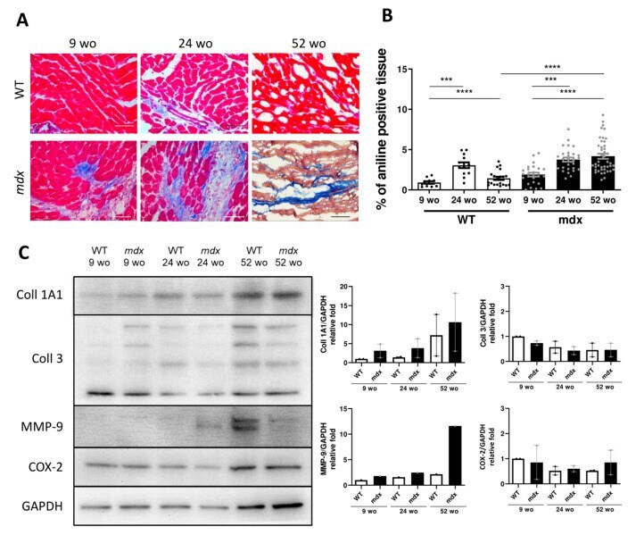
- Figure 4 Fibrotic deposit and cardiac dysfunction correlate with decreasing CVPC presence in mdx heart. ( A ) Representative images of histological analysis stained using Masson trichrome technique showing myocytes (in red) and collagenous fibrotic tissue (in blue) in the left ventricle of WT and mdx hearts at 9, 24, and 52 wo. Line represents 100 um. ( B ) The ratio of red and blue stained tissue was evaluated in WT hearts (open bars and black dots, n = 4-11 slices/3 animals per group) and mdx hearts (black bars and grey dots, n = 3-16 slices/3 animals per group) at the age of 24 wo and further at 52 wo. Statistical significance was calculated by Kruskal-Wallis test and Dunn's multiple comparison post-hoc test (*** p < 0.001, **** p < 0.0001). ( C ) Western blot analysis of collagen proteins and inflammatory proteins in the cardiac tissues. Left panel shows representative images of collagen 1A1 (Coll 1A1), collagen 3 (Coll 3), cyclooxygenase 2 (COX-2), and matrix metalloproteinase 9 (MMP-9) compared to the GAPDH control. The right panels show the normalized densitometry of each protein normalized by GAPDH content of WT (open bars, n = 2 animals) and mdx (black bars, n = 2 animals).
- Submitted by
- Invitrogen Antibodies (provider)
- Main image
- Experimental details
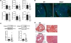
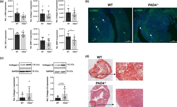
- FIGURE 2 Expression of fibrosis-related markers in PAD4 -/- hearts. (A) The expression of fibrosis-related genes in WT ( n = 7) and PAD4 -/- ( n = 10) hearts was analysed by real-time PCR. (B) alpha-SMA expression was detected by immunofluorescence and was found to be restricted to vascular cells ( white arrow heads ). One representative image of three stained hearts is depicted. Scale bar indicates 100 um. n = 5/group. (C) Collagen I and III protein expression in cardiac tissue of WT and PAD4 -/- mice. n = 7. (D) Representative images of heart sections from WT and PAD4-deficient mice stained with Masson trichrome under baseline conditions. * p < 0.05
- Submitted by
- Invitrogen Antibodies (provider)
- Main image
- Experimental details
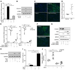
- Figure 6 FOXM1 inhibition prevents and reverses TGF-beta-induced myofibroblast differentiation and sensitizes myofibroblasts to FasL-induced apoptosis. ( A - C ) Effects of a 1-hour pretreatment of CCL210 cells with 2.5 muM Sio A (prevention protocol) on TGF-beta-induced expression of alpha-SMA mRNA ( A ) and protein ( B , Western blot; C , immunofluorescence microscopy) as well as collagen I protein ( B ). ( D ) Basal levels of ACTA2 mRNA in lung fibroblasts isolated from IPF patients or nonfibrotic controls ( n = 5). ( E ) Effect of transfection with FOXM1 siRNA or scrambled (control) siRNA (for 16 hours) (left) or 30-minute pretreatment with 2.5 muM Sio A (right) on ACTA2 mRNA levels in IPF or control lung fibroblasts ( n = 5) stimulated with and without TGF-beta for 24 hours. ( F ) Immunofluorescence microscopic analysis of alpha-SMA expression in TGF-beta-generated myofibroblasts treated with 2.5 muM Sio A for 24 hours (reversal protocol). ( G ) Effect of transfection with FOXM1 siRNA or scrambled (control) siRNA (for 16 hours) on FasL-induced apoptosis and expression of apoptosis-associated genes FAS , CASP3 , and BIRC5 by Western blot analysis of TGF-beta-generated myofibroblasts. ( H and I ) Effects of a 1-hour pretreatment with 2.5 muM Sio A on FasL-induced apoptosis in TGF-beta-generated myofibroblasts, as determined by cleaved PARP expression assessed by Western blot ( H ) and by the frequency of annexin V staining assessed by flow cytomet
