Explore
 Validate
 Learn
Learn Western blot
 ELISA
ELISAAntibody data
- Antibody Data
- Antigen structure
- References [2]
- Comments [0]
- Validations
- Western blot [2]
- Immunohistochemistry [3]
Submit
Validation data
Reference
Comment
Report error
- Product number
- R1038 - Provider product page

- Provider
- Acris Antibodies GmbH
- Proper citation
- Acris Antibodies GmbH Cat#R1038, RRID:AB_978381
- Product name
- anti Collagen type I
- Antibody type
- Polyclonal
- Antigen
- Collagen type I purified from Human and Bovine placenta.
- Reactivity
- Human, Mouse, Rat, Bovine
- Host
- Rabbit
- Vial size
- 0.1 mg
- Concentration
- 1.0 mg/ml (by UV absorbance at 280 nm)
Submitted references The epidermal basement membrane is a composite of separate laminin- or collagen IV-containing networks connected by aggregated perlecan, but not by nidogens.
Supramolecular interactions in the dermo-epidermal junction zone: anchoring fibril-collagen VII tightly binds to banded collagen fibrils.
Behrens DT, Villone D, Koch M, Brunner G, Sorokin L, Robenek H, Bruckner-Tuderman L, Bruckner P, Hansen U
The Journal of biological chemistry 2012 May 25;287(22):18700-9
The Journal of biological chemistry 2012 May 25;287(22):18700-9
Supramolecular interactions in the dermo-epidermal junction zone: anchoring fibril-collagen VII tightly binds to banded collagen fibrils.
Villone D, Fritsch A, Koch M, Bruckner-Tuderman L, Hansen U, Bruckner P
The Journal of biological chemistry 2008 Sep 5;283(36):24506-13
The Journal of biological chemistry 2008 Sep 5;283(36):24506-13
No comments: Submit comment
Supportive validation
- Submitted by
- Acris Antibodies GmbH (provider)
- Main image
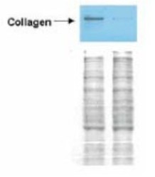
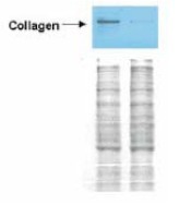
- Experimental details
- Western blot analysis is shown using Collagen type I antibody Cat.-No R1038 to detect expression of collagen I in Wistar rat hepatic stellate cells (HSC) in control (GFP-transduced) (left lane) and PPARgamma-transduced cell lysates (right lane). Protein staining shown below each blot depicts equal protein loading. An equal amount of the whole cell protein (100 µg) was separated by SDS-PAGE and electroblotted to nitrocellulose membranes. Proteins were detected by incubating the membrane with Collagen type I antibody at a concentration of 0.2-2 µg/10 ml in TBS (100 mM Tris-HCl, 0.15 M NaCl, pH 7.4) with 5% Non-fat milk. Detection occurred by incubation with a horseradish peroxidase-conjugated secondary antibody at 1 µg/10 ml. Proteins were detected by a chemiluminescent method using the PIERCE ECL kit (Amersham Biosciences). Other detection systems will yield similar results. See Hazra et al. (2004) for additional details.
- Submitted by
- Acris Antibodies GmbH (provider)
- Main image

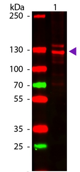
- Experimental details
- Western Blot of Human Collagen Type I using Collagen I Antibody (Cat.-No R1038). Lane 1: Human Collagen Type 1. Lane 2: None. Load: 50 ng per lane. Primary antibody: Collagen Type I antibody at 1:1,000 overnight at 4°C. Secondary antibody: DyLight⢠649 rabbit secondary antibody at 1:20,000 for 30 min at RT. Block: MB-070 for 30 min at RT. Predicted/Observed size: 139 & 130 kDa, 139 & 130 kDa for Collagen Type I. Other Band(s): Collagen Type I splice variants and isoforms.
Supportive validation
- Submitted by
- Acris Antibodies GmbH (provider)
- Main image
- Experimental details
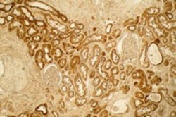
- Immunohistochemistry using affinity purified Collagen type I antibody Cat.-No R1038 at a 1/100 dilution to detect distal tubules in normal kidney tissue. Note the absence of staining of glomeruli. The antibody was reacted with antibody for 4 h RT followed by secondary antibody and substrate reaction. Tissue was Formalin-fixed and Paraffin embedded. No antigen retrieval was performed.

- Submitted by
- Acris Antibodies GmbH (provider)
- Main image
- Experimental details
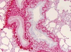
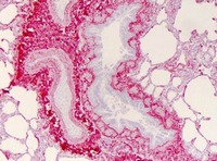
- Immunohistochemistry of human lung tissue (Formalin-fixed, Paraffin-embedded) using Collagen type I antibody Cat.-No R1038: Primary antibody (Collagen I) at 1:400, secondary antibody: Peroxidase goat anti-rabbit at 1:10,000 for 45 min at RT; Localization: Strong staining was observed in the extracellular matrix of the lung. Epithelial cells were negative; Staining: Antibody as precipitated red signal with a hematoxylin purple nuclear counterstain.
- Submitted by
- Acris Antibodies GmbH (provider)
- Main image
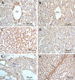
- Experimental details
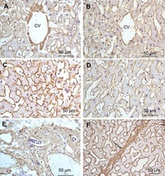
- Immunohistochemistry of a liver section (Formalin-fixed, Paraffin-embedded) using Collagen type I antibody Cat.-No R1038. A: Central vein (CV) fibrosis, B: Non-fibrotic CV, C: Perisinusodial fibrosis, D: Non-fibrotic area, E: Protat tract fibrosis, F: Septal fibrosis (arrow). Primary antibody: Collagen type I antibody at 1:1250 for 4°C for 24hr; Secondary antibody: Peroxidase biotin-streptavidin rabbit secondary antibody at 1:10,000 for 45 min at RT; Localization: Collagen type I is intra- and extracellular; Staining: 3.3â-diaminobenzidine tetrahydrochloride was used as the chromogen. Nuclei were counterstained purple with hematoxylin.