Explore
 Validate
 Learn
Learn Western blot
Antibody data
- Antibody Data
- Antigen structure
- References [0]
- Comments [0]
- Validations
- Western blot [2]
- Immunohistochemistry [1]
- Flow cytometry [1]
Submit
Validation data
Reference
Comment
Report error
- Product number
- PA5-47949 - Provider product page

- Provider
- Invitrogen Antibodies
- Product name
- BMPR2 Polyclonal Antibody
- Antibody type
- Polyclonal
- Antigen
- Recombinant full-length protein
- Description
- In direct ELISAs, less than 1% cross-reactivity with recombinant human (rh) BMPR-IA, recombinant mouse (rm) BMPR-IA, rhBMPR-IB, and rmBMPR-IB is observed. Reconstitute at 0.2 mg/mL in sterile PBS.
- Reactivity
- Human
- Host
- Goat
- Isotype
- IgG
- Vial size
- 100 µg
- Concentration
- 0.2 mg/mL
- Storage
- -20° C, Avoid Freeze/Thaw Cycles
No comments: Submit comment
Supportive validation
- Submitted by
- Invitrogen Antibodies (provider)
- Main image
- Experimental details
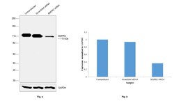
- Knockdown of BMPR2 was achieved by transfecting A-431 cells with BMPR2 specific siRNAs (Silencer® select Product # s2044). Western blot analysis (Fig. a) was performed using whole cell extracts from the BMPR2 knockdown cells (Lane 3), non-specific scrambled siRNA transfected cells (Lane 2) and untransfected cells (Lane 1). The blot was probed with Anti-BMPR2 Polyclonal Antibody (Product # PA5-47949, 0.1µg/ml) and Goat anti-Rabbit IgG (H+L), Superclonal™ Recombinant Secondary Antibody, HRP (Product # A27036, 1:4000 dilution) using the iBright FL 1000 (Product # A32752). Densitometric analysis of this Western Blot is shown in histogram (Fig. b). Decrease in signal upon siRNA mediated knock down confirms that antibody is specific to BMPR2.

- Submitted by
- Invitrogen Antibodies (provider)
- Main image
- Experimental details
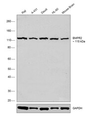
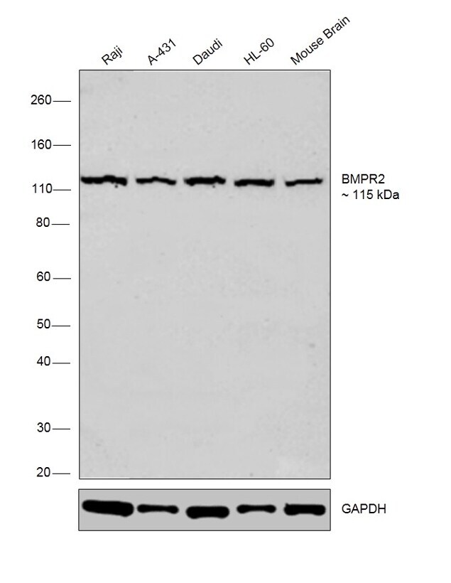
- Western blot was performed using Anti-BMPR2 Polyclonal Antibody (Product # PA5-47949) and a 115 kDa band corresponding to BMPR2 was observed across the cell lines and tissues tested. Whole cell extracts (30 µg lysate) of Raji (Lane 1), A-431 (Lane 2), Daudi (Lane 3), HL-60 (Lane 4) and tissue extract (30 µg lysate) of Mouse Brain (Lane 5) were electrophoresed using Novex® NuPAGE® 4-12 % Bis-Tris gel (Product # NP0322BOX). Resolved proteins were then transferred onto a nitrocellulose membrane (Product # IB23001) by iBlot® 2 Dry Blotting System (Product # IB21001). The blot was probed with the primary antibody (1:2000 dilution) and detected by chemiluminescence with Goat anti-Rabbit IgG (H+L), Superclonal™ Recombinant Secondary Antibody, HRP (Product # A27036, 1:4000 dilution) using the iBright FL 1000 (Product # A32752). Chemiluminescent detection was performed using Novex® ECL Chemiluminescent Substrate Reagent Kit (Product # WP20005).
Supportive validation
- Submitted by
- Invitrogen Antibodies (provider)
- Main image

- Experimental details
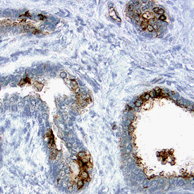
- Immunohistochemical analysis of BMPR2 in formalin fixed paraffin-embedded sections of human prostate. Samples were incubated in BMPR2 polyclonal antibody (Product # PA5-47949) using a dilution of 15 µg/mL overnight at 4 °C. Tissue was stained using the Anti-Goat HRP-DAB Cell & Tissue Staining Kit (brown) and counterstained with hematoxylin (blue). Specific labeling was localized to the plasma membrane of epithelial cells.
Supportive validation
- Submitted by
- Invitrogen Antibodies (provider)
- Main image

- Experimental details
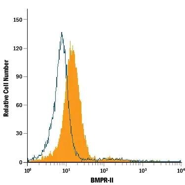
- Flow cytometry of BMPR2 in PC‚3 human prostate cancer cell line. Samples were incubated in BMPR2 polyclonal antibody (Product # PA5-47949) or control antibody followed by Allophycocyanin-conjugated Anti-Goat IgG Secondary Antibody.