 Explore
 Validate
 Learn
Learn Western blot
 Immunocytochemistry
Antibody data
- Antibody Data
- Antigen structure
- References [0]
- Comments [0]
- Validations
- Immunocytochemistry [3]
- Immunohistochemistry [3]
Submit
Validation data
Reference
Comment
Report error
- Product number
- GTX22804 - Provider product page

- Provider
- GeneTex
- Proper citation
- GeneTex Cat#GTX22804, RRID:AB_384861
- Product name
- AChR antibody [88B]
- Antibody type
- Monoclonal
- Reactivity
- Human, Mouse, Rat, Chicken/Avian, Rabbit
- Host
- Mouse
- Storage
- Keep as concentrated solution. Aliquot and store at -80?C or below. Avoid multiple freeze-thaw cycles.
No comments: Submit comment
Supportive validation
- Submitted by
- GeneTex (provider)
- Main image

- Experimental details
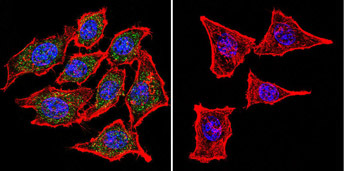
- Immunofluorescent analysis of Nicotinic Acetylcholine Receptor using Anti-Nicotinic Acetylcholine Receptor Monoclonal Antibody (88B) (GTX22804) shows staining in Hela Cells. Nicotinic Acetylcholine Receptor staining (green), F-Actin staining with Phalloidin (red) and nuclei with DAPI (blue) is shown. Cells were grown on chamber slides and fixed with formaldehyde prior to staining. Cells were probed without (control) or with or an antibody recognizing Nicotinic Acetylcholine Receptor (GTX22804) at a dilution of 1:100 over night at 4 C, washed with PBS and incubated with a DyLight-488 conjμgated secondary antibody.
- Submitted by
- GeneTex (provider)
- Main image
- Experimental details
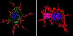
- Immunofluorescent analysis of Nicotinic Acetylcholine Receptor using Anti-Nicotinic Acetylcholine Receptor Monoclonal Antibody (88B) (GTX22804) shows staining in Neuro-2a Cells. Nicotinic Acetylcholine Receptor staining (green), F-Actin staining with Phalloidin (red) and nuclei with DAPI (blue) is shown. Cells were grown on chamber slides and fixed with formaldehyde prior to staining. Cells were probed without (control) or with or an antibody recognizing Nicotinic Acetylcholine Receptor (GTX22804) at a dilution of 1:100 over night at 4 C, washed with PBS and incubated with a DyLight-488 conjμgated secondary antibody.

- Submitted by
- GeneTex (provider)
- Main image

- Experimental details
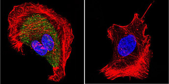
- Immunofluorescent analysis of Nicotinic Acetylcholine Receptor using Anti-Nicotinic Acetylcholine Receptor Monoclonal Antibody (88B) (GTX22804) shows staining in U251 Cells. Nicotinic Acetylcholine Receptor staining (green), F-Actin staining with Phalloidin (red) and nuclei with DAPI (blue) is shown. Cells were grown on chamber slides and fixed with formaldehyde prior to staining. Cells were probed without (control) or with or an antibody recognizing Nicotinic Acetylcholine Receptor (GTX22804) at a dilution of 1:20 over night at 4 C, washed with PBS and incubated with a DyLight-488 conjμgated secondary antibody.
Supportive validation
- Submitted by
- GeneTex (provider)
- Main image
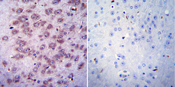
- Experimental details
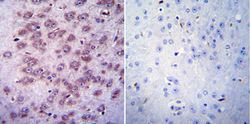
- Immunohistochemistry was performed on normal biopsies of de-paraffinized Mouse brain tissue. To expose target proteins, heat induced antigen retrieval was performed using 10mM sodium citrate (pH 6.0) buffer, microwaved for 8-15 minutes. Following antigen retrieval tissues were blocked in 3% BSA-PBS for 30 minutes at room temperature. Tissues were then probed at a dilution of 1:20 with a mouse monoclonal antibody recognizing Nicotinic Acetylcholine Receptor (GTX22804) or without primary antibody (negative control) overnight at 4°C in a humidified chamber. Tissues were washed extensively with PBST and endogenous peroxidase activity was quenched with a peroxidase suppressor. Detection was performed using a biotin-conjμgated secondary antibody and SA-HRP, followed by colorimetric detection using DAB. Tissues were counterstained with hematoxylin and prepped for mounting.
- Submitted by
- GeneTex (provider)
- Main image
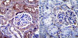
- Experimental details
- Immunohistochemistry was performed on normal biopsies of de-paraffinized Mouse kidney tissue. To expose target proteins, heat induced antigen retrieval was performed using 10mM sodium citrate (pH 6.0) buffer, microwaved for 8-15 minutes. Following antigen retrieval tissues were blocked in 3% BSA-PBS for 30 minutes at room temperature. Tissues were then probed at a dilution of 1:20 with a mouse monoclonal antibody recognizing Nicotinic Acetylcholine Receptor (GTX22804) or without primary antibody (negative control) overnight at 4°C in a humidified chamber. Tissues were washed extensively with PBST and endogenous peroxidase activity was quenched with a peroxidase suppressor. Detection was performed using a biotin-conjμgated secondary antibody and SA-HRP, followed by colorimetric detection using DAB. Tissues were counterstained with hematoxylin and prepped for mounting.

- Submitted by
- GeneTex (provider)
- Main image
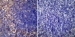
- Experimental details
- Immunohistochemistry was performed on normal biopsies of de-paraffinized mouse lymph node tissue. To expose target proteins, heat induced antigen retrieval was performed using 10mM sodium citrate (pH6.0) buffer, microwaved for 8-15 minutes. Following antigen retrieval tissues were blocked in 3% BSA-PBS for 30 minutes at room temperature. Tissues were then probed at a dilution of 1:20 with a mouse monoclonal antibody recognizing Nicotinic Acetylcholine Receptor (GTX22804) or without primary antibody (negative control) overnight at 4°C in a humidified chamber. Tissues were washed extensively with PBST and endogenous peroxidase activity was quenched with a peroxidase suppressor. Detection was performed using a biotin-conjμgated secondary antibody and SA-HRP, followed by colorimetric detection using DAB. Tissues were counterstained with hematoxylin and prepped for mounting.
