Explore
 Validate
 Learn
LearnPA5-47973
antibody from Invitrogen Antibodies
Targeting: GREM1
CKTSF1B1, CRAC1, DAND2, DRM, gremlin, HMPS
 Immunohistochemistry
 Blocking/Neutralizing
Antibody data
- Antibody Data
- Antigen structure
- References [1]
- Comments [0]
- Validations
- Immunohistochemistry [4]
- Other assay [1]
Submit
Validation data
Reference
Comment
Report error
- Product number
- PA5-47973 - Provider product page

- Provider
- Invitrogen Antibodies
- Product name
- GREM1 Polyclonal Antibody
- Antibody type
- Polyclonal
- Antigen
- Recombinant full-length protein
- Description
- In direct ELISAs, approximately 40% cross-reactivity with recombinant mouse (rm) PRDC/Gremlin-2 is observed, approximately 10% cross-reactivity with recombinant human (rh) Gremlin is observed and less than 5% cross-reactivity with rmDan, rhCerberus-1, and recombinant chicken Caronte is observed. Reconstitute at 0.2 mg/mL in sterile PBS. Endoxin level is
- Reactivity
- Human, Mouse
- Host
- Goat
- Isotype
- IgG
- Vial size
- 100 μg
- Concentration
- 0.2 mg/mL
- Storage
- -20°C, Avoid Freeze/Thaw Cycles
Submitted references Altered bone growth dynamics prefigure craniosynostosis in a zebrafish model of Saethre-Chotzen syndrome.
Teng CS, Ting MC, Farmer DT, Brockop M, Maxson RE, Crump JG
eLife 2018 Oct 25;7
eLife 2018 Oct 25;7
No comments: Submit comment
Supportive validation
- Submitted by
- Invitrogen Antibodies (provider)
- Main image

- Experimental details
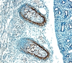
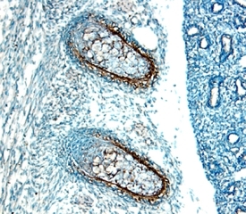
- Immunohistochemical analysis of GREM1 in immersion fixed frozen sections of embryonic mouse ribs (15 d.p.c.). Samples were incubated in GREM1 polyclonal antibody (Product # PA5-47973) using a dilution of 15 µg/mL overnight at 4 °C. Tissue was stained with the Anti-Goat HRP-DAB Cell & Tissue Staining Kit (brown) and counterstained with hematoxylin (blue).

- Submitted by
- Invitrogen Antibodies (provider)
- Main image

- Experimental details
- Immunohistochemical analysis of GREM1 in immersion fixed paraffin-embedded sections of human breast cancer tissue. Samples were incubated with GREM1 polyclonal antibody (Product # PA5-47973) using a dilution of 3 µg/mL for 1 hour at room temperature followed by Anti-Goat IgG VisUCyte™ HRP Polymer Antibody. Before incubation with the primary antibody, tissue was subjected to heat-induced epitope retrieval using Antigen Retrieval Reagent-Basic. Tissue was stained using DAB (brown) and counterstained with hematoxylin (blue). Specific staining was localized to cytoplasm.

- Submitted by
- Invitrogen Antibodies (provider)
- Main image
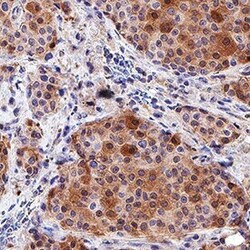
- Experimental details
- Immunohistochemical analysis of GREM1 in immersion fixed paraffin-embedded sections of human breast cancer tissue. Samples were incubated with GREM1 polyclonal antibody (Product # PA5-47973) using a dilution of 3 µg/mL for 1 hour at room temperature followed by Anti-Goat IgG VisUCyte™ HRP Polymer Antibody. Before incubation with the primary antibody, tissue was subjected to heat-induced epitope retrieval using Antigen Retrieval Reagent-Basic. Tissue was stained using DAB (brown) and counterstained with hematoxylin (blue). Specific staining was localized to cytoplasm.

- Submitted by
- Invitrogen Antibodies (provider)
- Main image
- Experimental details
- Immunohistochemical analysis of GREM1 in immersion fixed frozen sections of embryonic mouse ribs (15 d.p.c.). Samples were incubated in GREM1 polyclonal antibody (Product # PA5-47973) using a dilution of 15 µg/mL overnight at 4 °C. Tissue was stained with the Anti-Goat HRP-DAB Cell & Tissue Staining Kit (brown) and counterstained with hematoxylin (blue).
Supportive validation
- Submitted by
- Invitrogen Antibodies (provider)
- Main image
- Experimental details
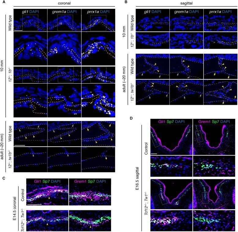
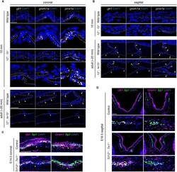
- Figure 7. Reduced osteoprogenitor pool at the mutant coronal suture. ( A, B ) Sections of forming coronal and sagittal sutures of 10 mm fish and fully formed sutures of adult fish were assessed for gli1 , grem1a , and prrx1a mRNA expression (white) by RNAscope in situ hybridization. Orange dotted lines indicate bones, and the boxed regions of the coronal suture regions are magnified below. For adult sutures, yellow arrowheads show expression in the suture mesenchyme, yellow arrows show expression of prrx1a in the periosteum, and grew arrows show lack of expression of gli1 and grem1a in the periosteum. Scale bar at 10 mm stage, 20 um; scale bar at adult stage, 50 um. ( C, D ) Sections of E14.5 coronal sutures and E16.5 forming sagittal sutures of mice stained for Gli1/Grem1 (magenta) and Sp7 protein (green). Yellow arrowheads indicate progenitor regions in forming coronal sutures. Boxed regions of parietal bone fronts in the forming sagittal sutures are magnified in lower panels. Nuclei are stained blue by DAPI in all images. Scale bars, 100 um. Figure 7--figure supplement 1. Quantification of progenitor marker and Fgf pathway transcripts in tcf12 ; twist1b mutants. ( A ) The number of gli1 , grem1a , and prrx1a transcripts at the forming coronal and sagittal suture regions were quantified per region area (between the bone fronts in Figure 7A ). Although the progenitor regions expressing these genes were reduced ( Figure 7B ), no significant d