 Explore
 Validate
 Learn
Learn Western blot
Antibody data
- Antibody Data
- Antigen structure
- References [0]
- Comments [0]
- Validations
- Western blot [3]
- Chromatin Immunoprecipitation [1]
- Other assay [1]
Submit
Validation data
Reference
Comment
Report error
- Product number
- PA5-30334 - Provider product page

- Provider
- Invitrogen Antibodies
- Product name
- SETDB1 Polyclonal Antibody
- Antibody type
- Polyclonal
- Antigen
- Recombinant protein fragment
- Description
- Recommended positive controls: NT2D1, IMR32, U87-MG, MCF-7.
- Concentration
- 1 mg/mL
No comments: Submit comment
Supportive validation
- Submitted by
- Invitrogen Antibodies (provider)
- Main image

- Experimental details
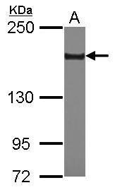
- Western Blot using SETDB1 Polyclonal Antibody (Product # PA5-30334). Sample (30 µg of whole cell lysate). Lane A: U87-MG. 5% SDS PAGE. SETDB1 Polyclonal Antibody (Product # PA5-30334) diluted at 1:2,000. The HRP-conjugated anti-rabbit IgG antibody was used to detect the primary antibody.
- Submitted by
- Invitrogen Antibodies (provider)
- Main image

- Experimental details
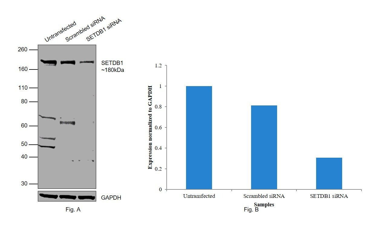
- Knockdown of SETDB1 was achieved by transfecting MCF7 with SETDB1 specific siRNAs (Silencer® select Products # s19111, s19110). Western blot analysis (Fig. a) was performed using modified whole cell extracts (1% SDS) from the SETDB1 knockdown cells (Lane 3), non-specific scrambled siRNA transfected cells (Lane 2) and untransfected cells (Lane 1). The blot was probed with SETDB1 Polyclonal Antibody (Product # PA5-30334, 1:1000 dilution) and Goat anti-Rabbit IgG (H+L) Superclonal™ Recombinant Secondary Antibody, HRP (Product # A27036, 1:4000 dilution). Densitometric analysis of this western blot is shown in histogram (Fig. b). Reduction in signal upon siRNA mediated knock down confirms that antibody is specific to SETDB1. Few uncharacterized bands were observed between 50-70 kDa.
- Submitted by
- Invitrogen Antibodies (provider)
- Main image
- Experimental details
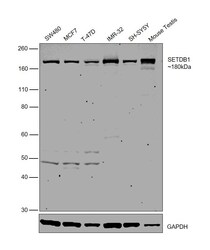
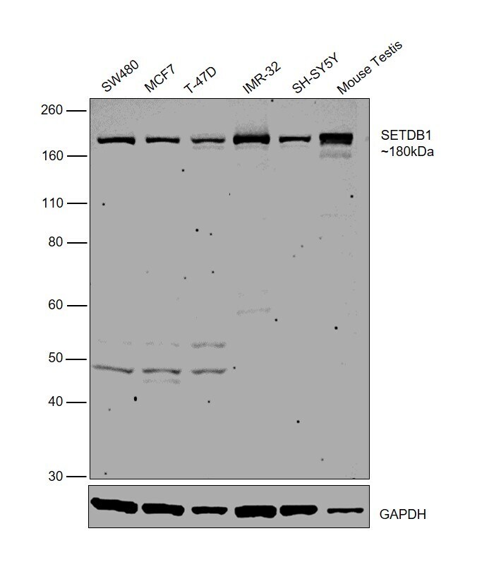
- Western blot was performed using Anti-SETDB1 Polyclonal Antibody (Product # PA5-30334) and a 180 kDa band corresponding to SETDB1 was observed in all tested cell and tissue lysates. Modified whole cell lysates (1% SDS) (30 µg lysate) of SW480 (Lane 1), MCF7 (Lane 2), T-47D (Lane 3), IMR-32 (Lane 4), SH-SY5Y (Lane 5), and tissue lysates (30 µg lysate) of Mouse Testis (Lane 6) were electrophoresed using Novex® NuPAGE® 4-12 % Bis-Tris gel (Product # NP0322BOX). Resolved proteins were then transferred onto a nitrocellulose membrane (Product # IB23001) by iBlot® 2 Dry Blotting System (Product # IB21001). The blot was probed with the primary antibody (1:1000 dilution) and detected by chemiluminescence with Goat anti-Rabbit IgG (H+L) Superclonal™ Recombinant Secondary Antibody, HRP (Product # A27036, 1:4000 dilution) using the iBright FL 1000 (Product # A32752). Chemiluminescent detection was performed using Novex® ECL Chemiluminescent Substrate Reagent Kit (Product # WP20005).
Supportive validation
- Submitted by
- Invitrogen Antibodies (provider)
- Main image
- Experimental details
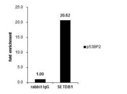
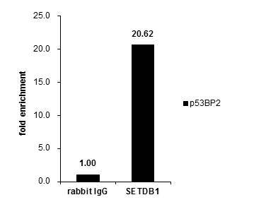
- Cross-linked ChIP was performed with HeLa chromatin extract and 5 µg of either control rabbit IgG or SETDB1 Polyclonal Antibody (Product # PA5-30334). The precipitated DNA was detected by PCR with primer set targeting to P53P2.
Supportive validation
- Submitted by
- Invitrogen Antibodies (provider)
- Main image
- Experimental details
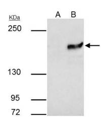
- SETDB1 Polyclonal Antibody immunoprecipitates SETDB1 protein in IP experiments. IP samples: HeLa nuclear extract. A. Control with 4 µg of preimmune Rabbit IgG. B. Immunoprecipitation of SETDB1 protein by 4 µg SETDB1 Polyclonal Antibody (Product # PA5-30334). 5 % SDS-PAGE. The immunoprecipitated SETDB1 protein was detected by SETDB1 Polyclonal Antibody (Product # PA5-30334) diluted at 1:1,000.
