 Explore
 Validate
 Learn
Learn Flow cytometry
Antibody data
- Antibody Data
- Antigen structure
- References [0]
- Comments [0]
- Validations
- Flow cytometry [2]
Submit
Validation data
Reference
Comment
Report error
- Product number
- FAB7738G - Provider product page

- Provider
- Novus Biologicals
- Product name
- Rat Monoclonal PD-1 Antibody
- Antibody type
- Monoclonal
- Description
- Protein A or G purified from hybridoma culture supernatant. Detects mouse PD-1 in direct ELISAs. In direct ELISAs, no cross-reactivity with recombinant human PD-1 is observed.
- Reactivity
- Mouse
- Host
- Rat
- Conjugate
- Green dye
- Isotype
- IgG
- Vial size
- 100 Tests
- Storage
- Protect from light. Do not freeze. 12 months from date of receipt, 2 to 8 degreesC as supplied.
No comments: Submit comment
Supportive validation
- Submitted by
- Novus Biologicals (provider)
- Main image

- Experimental details
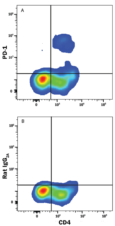
- Detection of PD-1 in Mouse Thymocytes by Flow Cytometry. Mouse thymocytes gated on CD3+ CD8+ cells were stained with Rat Anti-Mouse CD4 Alexa Fluor® 405-conjugated Monoclonal Antibody (Catalog # FAB554V) and either (A) Rat Anti-Mouse PD-1 Alexa Fluor® 488-conjugated Monoclonal Antibody (Catalog # FAB7738G) or (B) Rat IgG2A Alexa Fluor 488 Isotype Control (Catalog # IC006G). View our protocol for Staining Membrane-associated Proteins.
- Submitted by
- Novus Biologicals (provider)
- Main image

- Experimental details
- Detection of PD-1 in Mouse Splenocytes by Flow Cytometry. Mouse splenocytes either (A) resting or (B) treated with 5 μg/mL of PHA for 72 hours were stained with Rat Anti-Mouse PD-1 Alexa Fluor® 488-conjugated Monoclonal Antibody (Catalog # FAB7738G, filled histogram) or isotype control antibody (Catalog # IC006G, open histogram). View our protocol for Staining Membrane-associated Proteins.
