Explore
 Validate
 Learn
Learn Western blot
 Flow cytometry
Antibody data
- Antibody Data
- Antigen structure
- References [4]
- Comments [0]
- Validations
- Western blot [2]
- Immunohistochemistry [3]
Submit
Validation data
Reference
Comment
Report error
- Product number
- AF1021 - Provider product page

- Provider
- R&D Systems
- Product name
- Mouse PD-1 Antibody
- Antibody type
- Polyclonal
- Description
- Antigen Affinity-purified. Detects mouse PD-1 in direct ELISAs and Western blots. In direct ELISAs, approximately 20% cross-reactivity with recombinant human PD-1 is observed.
- Reactivity
- Mouse
- Host
- Goat
- Conjugate
- Unconjugated
- Antigen sequence
Q02242- Isotype
- IgG
- Vial size
- 100 ug
- Concentration
- LYOPH
- Storage
- Use a manual defrost freezer and avoid repeated freeze-thaw cycles. 12 months from date of receipt, -20 to -70 °C as supplied. 1 month, 2 to 8 °C under sterile conditions after reconstitution. 6 months, -20 to -70 °C under sterile conditions after reconstitution.
Submitted references IL-17C-mediated innate inflammation decreases the response to PD-1 blockade in a model of Kras-driven lung cancer.
CD8αα intraepithelial lymphocytes arise from two main thymic precursors.
Anti-programmed cell death 1 antibody reduces CD4+PD-1+ T cells and relieves the lupus-like nephritis of NZB/W F1 mice.
Depletion of the programmed death-1 receptor completely reverses established clonal anergy in CD4(+) T lymphocytes via an interleukin-2-dependent mechanism.
Ritzmann F, Jungnickel C, Vella G, Kamyschnikow A, Herr C, Li D, Menger MM, Angenendt A, Hoth M, Lis A, Bals R, Beisswenger C
Scientific reports 2019 Jul 17;9(1):10353
Scientific reports 2019 Jul 17;9(1):10353
CD8αα intraepithelial lymphocytes arise from two main thymic precursors.
Ruscher R, Kummer RL, Lee YJ, Jameson SC, Hogquist KA
Nature immunology 2017 Jul;18(7):771-779
Nature immunology 2017 Jul;18(7):771-779
Anti-programmed cell death 1 antibody reduces CD4+PD-1+ T cells and relieves the lupus-like nephritis of NZB/W F1 mice.
Kasagi S, Kawano S, Okazaki T, Honjo T, Morinobu A, Hatachi S, Shimatani K, Tanaka Y, Minato N, Kumagai S
Journal of immunology (Baltimore, Md. : 1950) 2010 Mar 1;184(5):2337-47
Journal of immunology (Baltimore, Md. : 1950) 2010 Mar 1;184(5):2337-47
Depletion of the programmed death-1 receptor completely reverses established clonal anergy in CD4(+) T lymphocytes via an interleukin-2-dependent mechanism.
Bishop KD, Harris JE, Mordes JP, Greiner DL, Rossini AA, Czech MP, Phillips NE
Cellular immunology 2009;256(1-2):86-91
Cellular immunology 2009;256(1-2):86-91
No comments: Submit comment
Supportive validation
- Submitted by
- R&D Systems (provider)
- Main image
- Experimental details
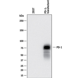
- Detection of Mouse PD-1 by Western Blot. Western blot shows lysates of 293T human embryonic kidney cell line mock transfected or transfected with mouse PD-1. PVDF membrane was probed with 0.2 µg/mL of Goat Anti-Mouse PD-1 Antigen Affinity-purified Polyclonal Antibody (Catalog # AF1021) followed by HRP-conjugated Anti-Goat IgG Secondary Antibody (Catalog # HAF017). A specific band was detected for PD-1 at approximately 75 kDa (as indicated). This experiment was conducted under reducing conditions and using Immunoblot Buffer Group 1.
- Submitted by
- R&D Systems (provider)
- Main image
- Experimental details
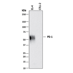
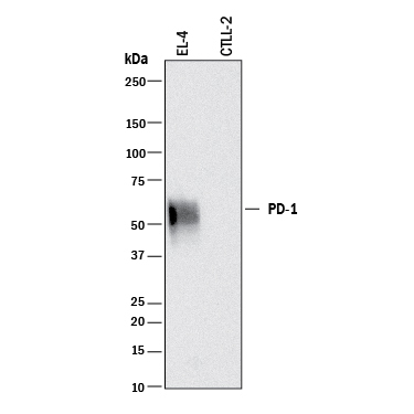
- Detection of Mouse PD-1 by Western Blot. Western blot shows lysates of EL-4 mouse lymphoblast cell line and CTLL-2 mouse cytotoxic T cell line (negative control). PVDF membrane was probed with 0.2 µg/mL of Goat Anti-Mouse PD-1 Antigen Affinity-purified Polyclonal Antibody (Catalog # AF1021) followed by HRP-conjugated Anti-Goat IgG Secondary Antibody (Catalog # HAF017). A specific band was detected for PD-1 at approximately 55 kDa (as indicated). This experiment was conducted under reducing conditions and using Immunoblot Buffer Group 1.
Supportive validation
- Submitted by
- R&D Systems (provider)
- Main image
- Experimental details
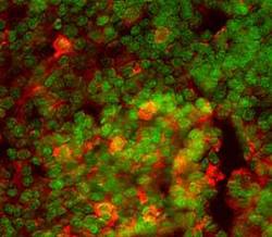
- PD-1 in Mouse Thymus. PD-1 was detected in perfusion fixed frozen sections of mouse thymus using 15 µg/mL Mouse PD-1 Antigen Affinity-purified Polyclonal Antibody (Catalog # AF1021) overnight at 4 °C. Tissue was stained (red) and counterstained (green). View our protocol for Fluorescent IHC Staining of Frozen Tissue Sections.

- Submitted by
- R&D Systems (provider)
- Main image
- Experimental details
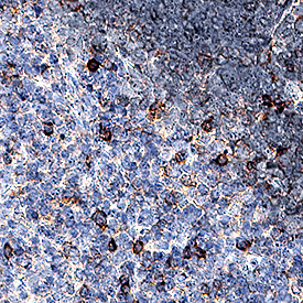
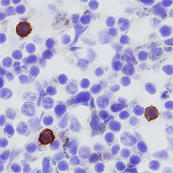
- PD-1 in Mouse Spleen. PD-1 was detected in perfusion fixed frozen sections of mouse spleen using Goat Anti-Mouse PD-1 Antigen Affinity-purified Polyclonal Antibody (Catalog # AF1021) at 5 µg/mL overnight at 4 °C. Tissue was stained using the Anti-Goat HRP-DAB Cell & Tissue Staining Kit (brown; Catalog # CTS008) and counterstained with hematoxylin (blue). Specific staining was localized to cytoplasm in splenocytes. View our protocol for Chromogenic IHC Staining of Frozen Tissue Sections.

- Submitted by
- R&D Systems (provider)
- Main image
- Experimental details
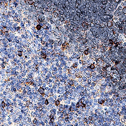
- PD-1 in Mouse Thymus. PD-1 was detected in perfusion fixed paraffin-embedded sections of mouse thymus using Goat Anti-Mouse PD-1 Antigen Affinity-purified Polyclonal Antibody (Catalog # AF1021) at 1.7 µg/mL for 1 hour at room temperature followed by incubation with the Anti-Goat IgG VisUCyte™ HRP Polymer Antibody (Catalog # VC004). Before incubation with the primary antibody, tissue was subjected to heat-induced epitope retrieval using Antigen Retrieval Reagent-Basic (Catalog # CTS013). Tissue was stained using DAB (brown) and counterstained with hematoxylin (blue). Specific staining was localized to cell membranes. View our protocol for IHC Staining with VisUCyte HRP Polymer Detection Reagents.